https://www.sciencedirect.com/science/article/pii/S2589004220301541
Tuore artikkeli, josta ilmenee KELCH ryhmän proteiini ja CUL5 CLR5 kompleksi säätelemässä SELENOS proteiinia. Artikkeli käsittelee KLHDC1, 2 ja 3 proteiinien suhdetta SELENOS proteiinin eri muotoihin proteiinin laadun kontrollissa.
- Cul5-type Ubiquitin Ligase KLHDC1 Contributes to the Elimination of Truncated SELENOS Produced by Failed UGA/Sec Decoding
Introduction
Protein
sequences generally consist of 20 common amino acids. The UGA mRNA
codon signals protein translation termination, but it can also be
translated into selenocysteine (Sec, U), the 21st amino acid, when a Sec
insertion sequence (SECIS) element in the mRNA 3′ untranslated region
(3′ UTR), Sec tRNA, Sec-specific elongation factor, and the
SECIS-binding protein SBP2 are present (Papp et al., 2007, Vindry et al., 2018).
At least 25 selenoproteins, including
- glutathione peroxidase,
- thioredoxin reductase, and
- selenoprotein S (SELENOS, formerly known as valosin-containing protein [VCP]-interacting membrane protein)
have been
identified (Davis et al., 2012, Gladyshev et al., 2016, Kryukov et al., 2003, Ye et al., 2004). Selenoproteins are largely involved in oxidation-reduction reactions (Davis et al., 2012).
In vitro,
SELENOS possesses both reductase and peroxidase activities, but only
when Sec is incorporated and not when Sec is mutated to Cys (Liu et al., 2013, Liu and Rozovsky, 2013). These findings indicate the importance of Sec for the oxidoreductase activity of SELENOS.
UGA redefinition is regulated by selenium availability (Huom: Dieettitekijä!) (Howard et al., 2013)
and involves a risk of premature translation termination. Indeed,
Sec-containing mature and Sec-lacking truncated selenoproteins have been
detected in human embryonic kidney (HEK)293 cells (Lin et al., 2015).
Human SELENOS consists of 189 amino acids, and Sec is the 188th amino
acid. Therefore, it is difficult to distinguish mature and truncated
SELENOS, and thus, it remains unknown how efficiently mature SELENOS is
translated. SELENOS is a single transmembrane protein located at the ER
membrane that eliminates misfolded proteins from the ER via
retro-translocation in collaboration with VCP (Christensen et al., 2012, Ye et al., 2004).
SELENOS expression is upregulated upon treatment with the ER
stress-inducer tunicamycin (TM), which prevents N-linked protein
glycosylation, and the knockdown of SELENOS inhibits membrane
translocation of VCP and enhances ER stress-induced apoptosis (Kim et al., 2007, Lee et al., 2014).
Importantly, Pro178 and Pro183, but not Sec188, are important for
interactions with VCP and ER-associated protein degradation (ERAD) (Lee et al., 2014).
The
two major functions of SELENOS, oxidoreductase activity and
ERAD-related functions, are dependent or independent on Sec,
respectively, indicating the importance of regulating the expression
levels of mature and truncated SELENOS.
(HUOM. SARS CoV ORF8 tekee interaktion CUL2, RBX1 ja ELOB kanssa)
Cullin 2 (Cul2)-containing
ubiquitin ligase has been shown to target truncated but not mature
SELENOS (Lin et al., 2015).
Cullin really interesting new gene (RING) ligases (CRLs) function in
the ubiquitin proteasome system, and a protein complex consisting of
Cul2, elongins B and C, von Hippel–Lindau (VHL) box protein, and
RING-box protein 1 (RBX1) belongs to the CRL superfamily (Kamura et al., 2004a, Lipkowitz and Weissman, 2011, Okumura et al., 2012).
Proteins containing the VHL box target their substrate for
ubiquitination and proteasomal degradation. Among VHL box proteins,
Kelch domain-containing (KLHDC) 2 and 3 have been shown to destabilize
truncated SELENOS directly or indirectly (Lin et al., 2015).
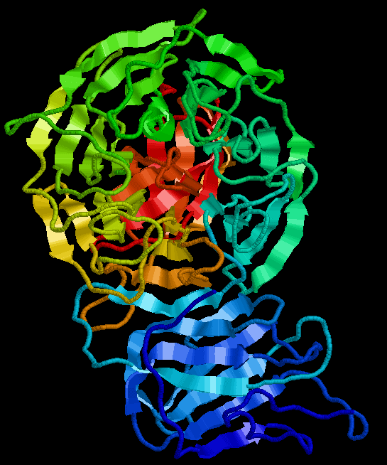
Five to seven Kelch repeats form a β-propeller structure that is involved in protein-protein interactions (Adams et al., 2000).
Recent research has revealed a “C-end rule,” which implies that the
amino acid sequence at the C terminus of a protein acts as a degron, a
specific, short peptide motif of a substrate protein recognized by
ubiquitin ligase (Koren et al., 2018, Lucas and Ciulli, 2017, Varshavsky, 1991).
KLHDC2 recognizes the C-terminal -Gly-Gly as a degron (Koren et al., 2018, Lin et al., 2018), whereas
KLHDC3 recognizes the C-terminal -Arg-(any amino acid, Xaa)n-Arg-Gly, -Arg-(Xaa)n-Lys-Gly, and -Arg-(Xaa)n-Gln-Gly sequences (Koren et al., 2018, Lin et al., 2018).
As the C terminus of mature SELENOS is -Gly-Gly-Sec-Gly, it is not
recognized by KLHDC2/3. However, truncated SELENOS ends with -Gly-Gly,
and thus, it is targeted by KLHDC2 for ubiquitination and proteasomal
degradation. KLHDC2 is the best-studied Cul2-type ubiquitin ligase that
targets truncated but not mature SELENOS.
KLHDC1, 2,
and 3 belong to the same protein family; their identity and similarity,
respectively, are as follows: 40.8% and 54.9% between KLHDC1 and KLHDC2,
23.1% and 34.2% between KLHDC1 and KLHDC3, and 23.5% and 36.7% between
KLHDC2 and KLHDC3 (based on pairwise alignments using EMBOSS Needle, https://www.ebi.ac.uk/Tools/psa/emboss_needle/).
Nevertheless, ectopic KLHDC1 and KLHDC2 predominantly localize to the cytoplasm and the nucleus, respectively (Chin et al., 2007),
suggesting that they have different biological roles. It has not clear
whether KLHDC1 also targets truncated SELENOS, as does KLHDC2, and
whether KLHDC1 also interacts with Cul2.
The aim of the current study
was to identify the substrate protein of KLHDC1. We purified and
identified KLHDC1-interacting proteins in HEK293T cells by mass
spectrometry. To study the hitherto unknown biological function of
KLHDC1, we established KLHDC1-knockdown U2OS cell lines and examined ER
stress-dependent cell death and reactive oxygen species (ROS) levels.
Results
KLHDC1 Interacts with SELENOS and Cul5
To
identify the potential KLHDC1-interacting proteins by mass
spectrometry, 3×FLAG-tagged KLHDC1 (3×FLAG-KLHDC1) was purified from
HEK293T cell lysates. Cul5, but not Cul2, and SELENOS were identified as
KLHDC1-interacting candidates. The peptides of SELENOS identified by
mass spectrometry are shown in Figure 1A.
To confirm this, 3×FLAG-KLHDC1, 3×FLAG-KLHDC2, and 3×FLAG-KLHDC3 were
expressed in HEK293T cells, cell lysates were subjected to
immunoprecipitation (IP) with an anti-FLAG antibody, and the
immunoprecipitates were analyzed by sodium dodecyl sulfate
polyacrylamide gel electrophoresis (SDS-PAGE) and immunoblotting (IB)
with an anti-SELENOS or anti-FLAG antibody. The interaction between
3×FLAG-KLHDC1 and endogenous SELENOS was confirmed (Figure 1B).
We also detected weak interaction between KLHDC2 and SELENOS. Given
that the anti-SELENOS antibody recognizes both mature and truncated
SELENOS, it was not clear which form is detected by KLHDC1. Next,
3×FLAG-KLHDC1, 3×FLAG-KLHDC2, and 3×FLAG-KLHDC3 were expressed in
HEK293T cells, cell lysates were subjected to IP with an anti-FLAG
antibody, and the immunoprecipitates were subjected to SDS-PAGE and IB
with an anti-Cul2, anti-Cul5, or anti-FLAG antibody. In line with
previously reported findings, KLHDC2 and KLHDC3 interacted with Cul2 (Koren et al., 2018, Lin et al., 2015, Lin et al., 2018) (Figure 1C). In contrast, KLHDC1 interacted only with Cul5, suggesting the existence of a Cul5 box, but not a Cul2 box, in this protein (Kamura et al., 2004b, Mahrour et al., 2008, Okumura et al., 2012).
The amino acid sequences of KLHDC1, KLHDC2, and KLHDC3 were aligned
with those of well-known Cul2 box-containing proteins including VHL
tumor suppressor (VHL), leucine-rich repeat protein (LRR)-1,
feminization 1 (FEM1)A, and preferentially expressed antigen of melanoma
(PRAME), as well as Cul5 box-containing proteins including
cytokine-inducible Src homology 2 domain-containing protein (CIS),
suppressor of cytokine signaling (SOCS)1, SOCS2, and SOCS3 (Figure 1D).
This revealed that KLHDC1 contains a consensus Cul5 box sequence
ϕxxLPϕPxxϕxxYL, whereas KLHDC2 and KLHDC3 contain the consensus Cul2 box
sequence ϕPxxϕxxxϕ, where ϕ represents hydrophobic amino acids and x
represents any amino acid. Next, we deleted the Cul5 box (amino acids
340–406) from KLHDC1 (KLHDC1(ΔCul5 box)), which resulted in a loss of
interaction with Cul5 (Figure 1E). These findings indicated that KLHDC1 is a Cul5-interacting protein that recognizes SELENOS.
Figure 1. KLHDC1 Interacts with SELENOS and Cul5
(A) Amino acid sequence of human SELENOS. Identified peptides by mass spectrometry are shown in bold font. U, selenocysteine.
(B)
SELENOS interacts with KLHDC1 and KLHDC2.
3×FLAG-KLHDC1, KLHDC2, or
KLHDC3 was expressed in HEK293T cells, and cell lysates were subjected
to immunoprecipitation (IP) with an anti-FLAG antibody and immunoblotted
with an anti-FLAG or anti-SELENOS antibody.
(C)
KLHDC1 is a Cul5-type ubiquitin ligase.
3×FLAG-KLHDC1, KLHDC2, or KLHDC3
was expressed in HEK293T cells, and cell lysates were subjected to IP
with an anti-FLAG antibody and immunoblotted with an anti-Cul2,
anti-Cul5, or anti-FLAG antibody.
(D) Amino acid
sequence alignment of Cul2 box- or Cul5 box-containing human proteins.
The numbers indicate the amino acid position in each protein. Conserved
Cul5 box and Cul2 box sequences are shown at the bottom or the top of
the box, respectively, in bold font. ϕ, hydrophobic residue.
(E)
Cul5 box-dependent interaction between KLHDC1 and Cul5.
3×FLAG-KLHDC1
(wild-type, WT) or a Cul5 box-deletion mutant KLHDC1(ΔCul5 box) was
expressed in HEK293T cells, and cell lysates were subjected to IP with
an anti-FLAG antibody and immunoblotted with an anti-Cul5 or anti-FLAG
antibody.
References
Lisätietoa KELCH proteiineista, joihin artikkelissa mainitut KLHDC proteiinit kuuluvat alaryhmänä. Kelch proteiineilla on propellirakenne. Kelch toistodomeenit muodostavat sen. Niitä on eri ryhmiä.
https://www.researchgate.net/publication/263432589/figure/fig1/AS:203157767495681@1425447994027/The-Kelch-Superfamily-A-The-Kelch-family-consists-of-63-proteins-that-are.png

https://en.wikipedia.org/wiki/Kelch_protein
https://www.researchgate.net/publication/263432589/figure/fig1/AS:203157767495681@1425447994027/The-Kelch-Superfamily-A-The-Kelch-family-consists-of-63-proteins-that-are.png
- Adams et al., 2000 J. Adams, R. Kelso, L. CooleyThe kelch repeat superfamily of proteins: propellers of cell function Trends Cell Biol., 10 (2000), pp. 17-24 Article Download PDFView Record in ScopusGoogle Schola Abstract
The kelch motif was discovered as a sixfold tandem element in the
sequence of the Drosophila kelch ORF1 protein. The repeated kelch motifs
predict a conserved tertiary structure, a β-propeller. This module
appears in many different polypeptide contexts and contains multiple
potential protein-protein contact sites. Members of this growing
superfamily are present throughout the cell and extracellularly and have
diverse activities. In this review, we discuss current information
concerning the structural organization of kelch repeat proteins, their
biological roles and the molecular basis of their action. © 2000
Elsevier Science Ltd.
Inga kommentarer:
Skicka en kommentar