Leta i den här bloggen

måndag 29 november 2021

Katsahdusta sars-2- viruksen S-proteiinin EFR (156-158)- alueen mutaatioihin. (Delta) E157G mutaatio S:NTD).

 https://www.frontiersin.org/articles/10.3389/fcimb.2021.765039/full

S-piikki:  E156G mutaation taustaa 

Sars-2 proteiinin  S-proteiinissa  N-terminaalisessa  domeenissa on kohta, kolme peräkkäistä aminohappoa E156, F157 ja R158 ja se  kohta on  muutoksille altista. Deltavirukselle näyttää olevan tyypillistä tehdä siihen kohtaan deleetio niin että F ja R poistuvat ja E-kohta taas saa substituution. Siihen kohtaan tulee G. 
Tällaisen muutoksen syntymisjärjestystä katson tänään, sillä papereistani huomaan maininnan, että jossain vaiheessa on ollut  R158G-muoto olemassa. Tuo ylläoleva linkki osoittaakin, että se on varhaisinta tapahtumaa tässä deltaviruksessa. Mutta sitten ei olekaan  viruksen RBD-motiivissa( Receptor Binding Motif) enää tällaista  substituutiota havaittavissa. Sen sijaan deltojen ominaispiirteissä mainitaan mutaatio E156G ja "deleetio 157-158".  Tuo varhaisin  mutaatio oli ohimenevä ja kaikesta tästä  "EFR"- kohdasta  on jäljellä vain  "G". kaikissa deltoissa ja sen varianttilaumassa , tosin jokaisen  yli 120 variantin  piikkiproteiinihahmoa en ole vielä  kirjannut vihkooni. 

Onko tällä EFR- alueen  deleetio-mutaatiolla merkitystä Sars-2-viruksessa? 
 
Asiaa on tutkittu esim   deltan lähisukulaisesta B.1.617.3:
 
 
 B.1.617.3 SARS CoV-2 spike E156G/Δ157-158 mutations contribute to reduced neutralization  sensitivity and increased infectivity.

Sitaatti:
"We found a total of eight changes; three were in NTD (N- terminal domain), four in RBD (receptor binding domain), and one on the S2 portion of ICS-05 spike (Figure-1a). Interestingly, we observed a six-nucleotide deletion that resulted in the loss of two amino acids at 157 and 158 positions and a
change of Glutamic acid at 156 positions to glycine (E156G/Δ157-158) (Supplementary figure-1).
This deletion we found in five of the total seven spike sequences isolated from the RT-PCR positive  cases (Supplementary figure-1). Out of these five cases, two were fully vaccinated. When the ICS-05 spike sequence was aligned with the available spike sequences on GISAID15, it corresponded to the  B.1.617 lineage (parent) , specifically with B.1.617.3 of the delta variant of concern (Figure-1b). Surfaced in  the month of March 2021, the delta variant dominated the second wave in the country (Figure-1c)  and was reported to have caused 25.3% of breakthrough infections 5
 
The E156G/∆157-158 mutation, first detected on 7th August 2020, subsequently became 35% prevalent worldwide (Figure-1d), and by August 2021, it was found in more than 85% of reported sequences from the USA, UK, and India (Figure-1f). 
The E156G/∆157-158 mutation has been detected with high frequency (Figure-1e) in at least 157 countries and is found in multiple PANGO lineages (Abdel Latif 2021; Elbe  and Buckland-Mer re tt, 2017). Given the higher prevalence worldwide and in the most affected  countries (Figure-1d, 1f), we hypothesized a functional relevance of E156G/Δ157-158 mutations. 
 

 Abstraktista näkyy vielä toisiakin  mutaatioita jotka vahvistavat  viruksen   kykyjä: 

Abstract
SARS CoV-2 variants raise significant concerns due to their ability to cause vaccine breakthrough  infections. Here, we sequence-characterized the spike gene, isolated from a breakthrough infection,  that corresponded to B.1.617.3 lineage. Delineating the functional impact of spike mutations using
reporter pseudoviruses (PV) revealed that N-terminal domain (NTD)-specific E156G/Δ157-158  contributed to increased infectivity and reduced sensitivity to ChAdOx1 nCoV-19 vaccine  (CovishieldTM)-elicited neutralizing antibodies. A six-nucleotide(nt)  deletion (467-472) in the spike
coding region introduced this change in the NTD. 

We confirmed the presence of E156G/Δ157-158  in the RT-PCR-positive cases concurrently screened, in addition to other circulating spike (S1) mutations like T19R, T95I, L452R, E484Q, and D614G. 

Notably, E156G/Δ157-158 was present in  more than 85% of the sequences reported from the USA, UK, and India in August 2021. The spike PV(pseudovirus) bearing combination of E156G/Δ157-158 and L452R further promoted infectivity and  conferred immune evasion

Additionally, increased cell-to-cell fusion was observed when spike
harbored E156G/Δ157-158, L452R, and E484Q, suggesting a combinatorial effect of these mutations.

 Notwithstanding, the plasma from a recovered individual robustly inhibited mutant spike  PV, indicating the increased breadth of neutralization post-recovery. Our data highlights the  importance of spike NTD-specific changes in determining infectivity and immune escape of  variants.


Keywords: E156G/Δ157-158 spike protein; breakthrough infection; cell-to-cell fusion; Spike NTD;  ChAdOx1 nCOV-19 vaccine

On mahdollista löytää myös  F157S vaihtoehtoinen mutaatio. 

B.1.526.1.

lördag 27 november 2021

Virusmutaatioiden heijastuma Sars-2 virusproteiineihin. Turkkilainen artikkeli valaisee asiaa.

https://onlinelibrary.wiley.com/doi/10.1002/jmv.27188 

Tässä artikkelissa mainitaan  muutoksista, jotka ovat taustalla  nsp12-virusproteiinin ja Sars-2 S-piikin mutaatioissa. Myös muitakni mutaatioita  esitetään. N-proteiinin, ja nsp3 proteiinin  mutaatioita emerkiksi.

Kirjoittaja mainitsee  löytämiään uusi mtaatioita.

fredag 26 november 2021

B.1.1 linjasta kehittynyt B.1.1.529 on lokiteltu Omicroniksi WHO-nimeltään. vastikään


Classification of Omicron (B.1.1.529): SARS-CoV-2 Variant of Concern

26 November 2021
Statement
Reading time: 2 min (616 words)

The Technical Advisory Group on SARS-CoV-2 Virus Evolution (TAG-VE) is an independent group of experts that periodically monitors and evaluates the evolution of SARS-CoV-2 and assesses if specific mutations and combinations of mutations alter the behaviour of the virus. The TAG-VE was convened on 26 November 2021 to assess the SARS-CoV-2 variant: B.1.1.529.

The B.1.1.529 variant was first reported to WHO from South Africa on 24 November 2021. The epidemiological situation in South Africa has been characterized by three distinct peaks in reported cases, the latest of which was predominantly the Delta variant. In recent weeks, infections have increased steeply, coinciding with the detection of B.1.1.529 variant. The first known confirmed B.1.1.529 infection was from a specimen collected on 9 November 2021.

This variant has a large number of mutations, some of which are concerning. Preliminary evidence suggests an increased risk of reinfection with this variant, as compared to other VOCs. The number of cases of this variant appears to be increasing in almost all provinces in South Africa. Current SARS-CoV-2 PCR diagnostics continue to detect this variant. Several labs have indicated that for one widely used PCR test, one of the three target genes is not detected (called S gene dropout or S gene target failure) and this test can therefore be used as marker for this variant, pending sequencing confirmation. Using this approach, this variant has been detected at faster rates than previous surges in infection, suggesting that this variant may have a growth advantage.

There are a number of studies underway and the TAG-VE will continue to evaluate this variant. WHO will communicate new findings with Member States and to the public as needed.

Based on the evidence presented indicative of a detrimental change in COVID-19 epidemiology, the TAG-VE has advised WHO that this variant should be designated as a VOC, and the WHO has designated B.1.1.529 as a VOC, named Omicron.

As such, countries are asked to do the following:

  • enhance surveillance and sequencing efforts to better understand circulating SARS-CoV-2 variants.
  • submit complete genome sequences and associated metadata to a publicly available database, such as GISAID.
  • report initial cases/clusters associated with VOC infection to WHO through the IHR mechanism.
  • where capacity exists and in coordination with the international community, perform field investigations and laboratory assessments to improve understanding of the potential impacts of the VOC on COVID-19 epidemiology, severity, effectiveness of public health and social measures, diagnostic methods, immune responses, antibody neutralization, or other relevant characteristics.

Individuals are reminded to take measures to reduce their risk of COVID-19, including proven public health and social measures such as wearing well-fitting masks, hand hygiene, physical distancing, improving ventilation of indoor spaces, avoiding crowded spaces, and getting vaccinated.

For reference, WHO has working definitions for SARS-CoV-2 Variant of Interest (VOI) and Variant of Concern (VOC).

A SARS-CoV-2 VOI is a SARS-CoV-2 variant:

  • with genetic changes that are predicted or known to affect virus characteristics such as transmissibility, disease severity, immune escape, diagnostic or therapeutic escape; AND
  • that has been identified as causing significant community transmission or multiple COVID-19 clusters, in multiple countries with increasing relative prevalence alongside increasing number of cases over time, or other apparent epidemiological impacts to suggest an emerging risk to global public health. 

A SARS-CoV-2 VOC is a SARS-CoV-2 variant that meets the definition of a VOI (see above) and, through a comparative assessment, has been demonstrated to be associated with one or more of the following changes at a degree of global public health significance:

  • increase in transmissibility or detrimental change in COVID-19 epidemiology; OR
  • increase in virulence or change in clinical disease presentation; OR
  • decrease in effectiveness of public health and social measures or available diagnostics, vaccines, therapeutics


Pohdintaa C.1.2 Sars Cov-2 variantista ja sen alainjoista


SARS-COV-2 C.1.2 variant is highly mutated but may possess reduced affinity for ACE2 receptor

Xiang-Jiao Yang
view affiliations
bioRxiv
published 19 October 2021 accessed 20 October 2021
Record provided by bioRxivdata.curatedBy.nameLearn more
SARS-COV-2 evolution generates different variants and drives the pandemic. As the current main driver, delta variant bears little resemblance to the other three variants of concern (alpha, beta and gamma), raising the question what features the future variants of concern may possess. To address this important question, I searched through the GISAID database for potential clues. While investigating how beta variant has been evolving in South Africa, I noticed a small group of genomes mainly classified as C.1.2 variant, with one-year old boy identified in March 2021 being the index case. Over 80% patients are younger than 60. At the average, there are 46-47 mutations per genome, making this variant one of the most mutated lineages identified. A signature substitution is spike Y449H. Like beta and gamma variants, C.1.2 possesses E484K and N501Y. The genomes are heterogenous and encode different subvariants. Like alpha variant, one such subvariant encodes the spike substitution P681H at the furin cleavage site. In a related genome, this substitution is replaced by P681R, which is present in delta variant. In addition, similar to this variant of concern, three C.1.2 subvariants also encode T478K. Mechanistically, spike Y449 recognizes two key residues of the cell-entry receptor ACE2 and Y449H is known to impair the binding to ACE2 receptor, so C.1.2 variant may show reduced affinity for this receptor. If so, this variant needs other mutations to compensate for such deficiency. These results raise the question whether C.1.2 variant is as virulent as suggested by its unexpected high number of mutations.
Funder

Kolumbiassa on predominanttia Mu varianttia. Artikkeli.

Kolumbian tilanteesta löytyy  netistä artikkeli:  Siell on koronaan sairastuneita n 5 miljoonaa ja menehtyneitä  yli 120 000, joten kuolleisuus  sairastuneista on  n 2.4  % prosentin luokkaa. Sitaatti:

 https://news.un.org/en/story/2021/09/1098942

A new coronavirus “variant of interest” named Mu – also known by its scientific name as B.1.621 – is being closely monitored by the World Health Organization (WHO), the agency has said.

In its weekly epidemiological update, published on Tuesday, WHO warned it was becoming increasingly prevalent in Colombia and Ecuador, and showed signs of possible resistance to vaccines.

Mu was first identified in Colombia in January 2021, and since then, there have been “sporadic reports” of cases and outbreaks in South America and Europe, WHO said.

While the global prevalence of Mu among sequenced COVID-19 cases is below 0.1%, its prevalence has “consistently increased” in Colombia and Ecuador, where it is now responsible for around 39% and 13% of infections, respectively.

Reports on the variant’s prevalence should be “interpreted with due consideration” given the low sequencing capacity of most countries, the agency said. 

More resistant to vaccines

Mu is the fifth variant of interest (VOI) to be monitored by the WHO since March. It has a number of mutations that suggest it could be more resistant to vaccines, the health agency warned, but stressed that further research would be needed to confirm this.

Preliminary data show a reduced effectiveness of vaccines “similar to that seen for the Beta variant”. The WHO said it would be monitoring “the epidemiology of the Mu variant in South America, particularly with the co-circulation of the Delta variant…for changes”

More infections

As of 29 August, over 4,500 sequences (3,794 B.1.621 sequences and 856 B.1.621.1 sequences), genome sequences, analysed samples of the virus taken from patients, have been designated as Mu in the past four weeks. The sequences are used to track how it moves through the population, on an open-source genome repository, known as GISAID.

Most of these have been reported in the U.S (2,065) and Colombia (852), Mexico (357) and Spain (473). 

Although this figure will be affected by both sequencing capacity, surveillance and the total number of cases in an area.

The novel coronavirus pandemic has killed at least 124,811 people in Colombia, according to a WHO report on Wednesday.                ( Artikkeli julkaistu 1 syyskuuta 2021)

More than 4,905,258 confirmed cases have been officially diagnosed across the country since the start of the epidemic. As of August 27, a total of 34,247,170 doses of vaccine had been administered.

C.1.2, not currently a concern

South African scientists are closely monitoring the development of another new variant there, according to news reports in the country.  (PANGO B.1.1.1.1.2)

However, C.1.2, is not yet a variant to follow, nor a variant of concern, according to the classification of the World Health Organization.

"It does not appear that its circulation is increasing”, said Dr. Margaret Harris, a spokesperson for the WHO, during a UN press briefing in Geneva on Tuesday.

 (KOSKA artikkeli on syyskun alusta, katson Kolumbian koronatilanteen nyt 3 kuukauden jälkeen WHO lähteestä). 

 https://covid19.who.int/region/amro/country/coTämän käyrän mukaan kuolleisuus seuraa tarttuuvuuden käyrän muotoa n. 2.4 prosenttina . Syyskuun jälkeen käyrä on laskenut phjiin. altoilun jälkeen. Viimeinen aalto li kaksikyttyräinen enen laskuaan nykyaallonphjaan. 


Katsausta kumulatiivisen prevalenssin prosenttilukuihin sars-2 virusvarianttien ilmenemisessä

 Olen summittain ja harpoten  käymässä läpi WHO lähteestä käyriä, joista havainnoituu sars-2  viruksen relevanssi  maailmassa. Ajallisesti käyrät kattavat  pandemian ajan kulloinkin   kyseessä olevan  virusvariantin osalta  Käytär ovat suhteellisia , koska  toisia viruksia esiintyy vain muutama sata ja toisia kymmenin tuhansin, joten täytyy tarkistaa  y-akselisa  missä prosenttilukemassa ( yksikön osalta)  milloinkin ollaan: 0,01 , 0,1%, 1 % vai 100% suuruusluokka y-akselilla .

En ole vielä päässyt kaikkien  deltavarianttien (AY linjojen)  käyriä kirjoittamaan vihkooni. Niitä on paljon , toista sataa. Katsoin välillä Alfan , Beetan, Gamman, Lambdan ja alalinjojen  käyrät ja lopuksi Mu variantin.

Mu -variantti yllätti, koska siinä on  merkitsevästi  poikkeavuutta muihin S-piikissä. Esim  yksi deleetiokohta (yy del145/144) on saanut subtituutionsa ja  siinä on nytY145N ja Y144S aminohappomuutokset . En kai ole tätä nähnyt muissa katsomissani  varianteissa.

Huomaan että Somessakin on  havaittu kolme Mu varianttia. Kolumbiassa niitä on havaittu 3720. Kaikenkaikkiaan niitä on havaittu 11 352 maailmassa. Sen esiintymisn korkein piikki (1%) on ollut keskikesällä.

 Siitä on  huomautuksia: https://outbreak.info/resources/2021.09.06.459005

https://outbreak.info/resources/2021.09.06.459005

Publication Preprint

Ineffective neutralization of the SARS-CoV-2 Mu variant by convalescent and vaccine sera

Keiya Uriu
Izumi Kimura
Kotaro Shirakawa
Akifumi Takaori-Kondo
Taka-aki Nakada
Atsushi Kaneda
- The Genotype to Phenotype Japan (G2P-Japan) Consortium
So Nakagawa, and 
Kei Sato
 
published 07 September 2021 accessed 20 October 2021
Record provided by bioRxivdata.curatedBy.nameLearn more
On August 30, 2021, the WHO classified the SARS-CoV-2 Mu variant (B.1.621 lineage) as a new variant of interest. The WHO defines "comparative assessment of virus characteristics and public health risks" as primary action in response to the emergence of new SARS-CoV-2 variants. Here, we demonstrate that the Mu variant is highly resistant to sera from COVID-19 convalescents and BNT162b2-vaccinated individuals. Direct comparison of different SARS-CoV-2 spike proteins revealed that Mu spike is more resistant to serum-mediated neutralization than all other currently recognized variants of interest (VOI) and concern (VOC). This includes the Beta variant (B.1.351) that has been suggested to represent the most resistant variant to convalescent and vaccinated sera to date (e.g., Collier et al, Nature, 2021; Wang et al, Nature, 2021). Since breakthrough infection by newly emerging variants is a major concern during the current COVID-19 pandemic (Bergwerk et al., NEJM, 2021), we believe that our findings are of significant public health interest. Our results will help to better assess the risk posed by the Mu variant for vaccinated, previously infected and naive populations.


måndag 22 november 2021

Galektiinin kaltainen alue Sars-S-proteiinin kärjissä

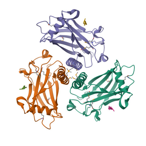
https://www.ebi.ac.uk/interpro/entry/InterPro/IPR032500/ 

hort name: bCoV_S1_N

Overlapping homologous superfamilies

domain relationships

Description

This entry represents the N-terminal domain of the betacoronavirus-like trimeric spike glycoprotein. The distal S1 subunit of the coronavirus spike protein is responsible for receptor binding. S1 contains two domains; an N-terminal galectin-like domain (NTD) and a receptor-binding domain (S1 RBD) also referred to as the S1 CTD or domain B. Either the S1 NTD or S1 RBD, or occasionally both, are involved in binding the host receptors. S1 NTD is located on the side of the spike trimer and mainly recognises sugar receptors
[1, 2, 3, 4]
. For many betacoronaviruses (bCoVs), for example mouse hepatitis virus (MHV), the RBD is located in the NTD. The structure of the MHV S1 NTD showed the same fold as human galectins (galactose-binding lectin), however it does not bind any sugar; instead, it binds to the carcinoembryonic antigen cell-adhesion molecule (CEACAM1) through protein-protein interactions
[2]
. All three CEACAM21a-binding sites in MHV spikes can be fully occupied by CEACAM1a. It has been shown that CEACAM1a binding to the MHV spike weakens the interactions between S1 and S2 and facilitates the proteolysis of the spike protein and dissociation of S1
[2]
. The homologous bovine CoV (BCov) S1 NTD also possesses a galectin fold but binds to sialic acid-containing moieties on host cell membranes, as does the NTD of three other group A b-Covs, namely human CoV (HCoV) OC43, avian b-CoV, and infectious bronchitis virus (IBV)
[5]
. Despite the S1 NTD of human respiratory b-CoV HKU1 being highly homologous to the NTDs of MHV and bovine CoV, it does not bind to either sugar or human CEACAMs and the RBD is found instead in the S1 RBD domain
[5]

The bCoV NTDs contain a conserved beta-sandwich core, but exhibit variant folds in the peripheral elements located in the top-ceiling region and on the lateral side. The core sandwich comprises in total sixteen anti-parallel beta-strands, assembling into three (upper, middle, and lower) beta-sheet layers. While showing different compositions and structures, the peripheral elements are topologically equivalent beta-sandwich-core insertions, highlighting a divergent evolution process for bCoVs to form different lineages
[3]
.

 Galektiini inhibiittorit, onko niistä sars-2 vastaisiksi lääkkeiksi?

file:///C:/Users/lea/AppData/Local/Temp/biomedicines-09-01208.pdf

 Abstract: Galectin-3 is a carbohydrate-binding protein and the most studied member of the galectin
family. It regulates several functions throughout the body, among which are inflammation and post-
injury remodelling. Recent studies have highlighted the similarity between Galectin-3s carbohydrate
recognition domain and the so-called “galectin fold” present on the N-terminal domain of the S1
sub-unit of the SARS-CoV-2 spike protein. Sialic acids binding to the N-terminal domain of the
Spike protein are known to be crucial for viral entry into humans, and the role of Galectin-3 as a
mediator of lung fibrosis has long been the object of study since its levels have been found to be
abnormally high in alveolar macrophages following lung injury. In this context, the discovery of
a double inhibitor may both prevent viral entry and reduce post-infection pulmonary fibrosis. In
this study, we use a database of 56 compounds, among which 37 have known experimental affinity
with Galectin-3. We carry out virtual screening of this database with respect to Galectin-3 and Spike
protein. Several ligands are found to exhibit promising binding affinity and interaction with the
Spike protein’s N-terminal domain as well as with Galectin-3. This finding strongly suggests that
existing Galectin-3 inhibitors possess dual-binding capabilities to disrupt Spike–ACE2 interactions.
Herein we identify the most promising inhibitors of Galectin-3 and Spike proteins, of which five
emerge as potential dual effective inhibitors. Our preliminary results warrant further in vitro and
in vivo testing of these putative inhibitors against SARS-CoV-2 with the hope of being able to halt
the spread of the virus in the future.
Keywords: Galectin-3; spike; dual inhibitors; COVID-19; docking

 1.1. Spike: Main Features and Functions
The Spike protein (also called S-protein) component of SARS-CoV-2 is a central target
in the fight against COVID-19 since it is the primary target of antibodies that provide
immunity against the virus. The surfaces of coronaviruses are covered with these spikes,
giving them their distinctive crown-like appearance in electron micrographs. The spikes
initiate the process of infection, binding to receptors and then fusing with the cell membrane to release the viral genome inside. Many other enveloped viruses, including influenza hemagglutinin and the envelope glycoproteins of HIV-1 and Ebola, use similar spike-likeproteins to infect cells.

 The spike protein is composed of three identical chains that together form a complex with a small domain inside the virus, a membrane-spanning segment,
and a large ectodomain that extends outward from the virus. 

 The S-protein exists in a meta stable prefusion conformation that undergoes a substantial structural rearrangement
to fuse the viral membrane with the host cell membrane. 

This process is triggered when the S1 sub-unit binds to a host cell receptor. Receptor binding destabilises the prefusion trimer, resulting in the shedding of the S1 sub-unit and transition of the S2 sub-unit to a stable postfusion conformation. 

To engage a host cell receptor, the receptor-binding domain (RBD)
of S1 undergoes hinge-like conformational movements that transiently hide or expose
the determinants of receptor binding. These two states are referred to as the “down” (or
“closed”) conformation and the “up” (or “open”) conformation (Figure 1), where down
corresponds to the receptor-inaccessible state and up corresponds to the receptor accessible state, which is presumed to be less stable [5].
The S1 sub-unit of the spike protein of SARS-CoV-2, critical for its entry into host
cells, can be subdivided into an NTD and a C-terminal domain (CTD) (Figure 2). 

 The
role of the CTD in viral entry has been well characterised in the literature, as it binds
angiotensin-converting-enzyme 2 (ACE2) receptors.
ACE2 is an enzyme that activates angiotensin, a peptide hormone involved in the
control of blood pressure. ACE2 is found on lung, heart, kidney, and intestinal cells, making these cells the primary targets for infection by the virus [5,6]. In addition, the spike is a glycoprotein: the ectodomain is covered with sugar chains that help to mask the virus from the immune system [6]. Watanabe et al. reported 22 N linked glycosylation sites on
Spike protein. In their study, across the 22 N-linked glycosylation sites, 52% were found to be fucosylated, while 15% of the glycans contained at least one sialic acid residue. Sialic acids, it should be noted, have been described as “eccentric” in that they bind several different pathogens and toxins. Owing to their location and abundant distribution, sialic acids participate in a wide variety of physiological and pathological processes. Sialic

 acids are a common constituent of glycoproteins, glycolipids, and gangliosides. They

Biomedicines 2021, 9, 1208 3 of 27
decorate the terminal of sugar chains at the surface of cells or soluble proteins. 

Sialic acids
linked to glycoproteins and gangliosides are recruited by a broad spectrum of viruses—
including coronaviruses—as receptors and/or attachment factors for cell entry. In many
viral infections (influenza, Ebola, SARSCoV, among others), glycan-mediated interactions are essential for the initial contact between the virus and the host [7–9]. The S1-NTD of SARS-CoV-2 has been shown to possess a sialic acid binding site highly similar to thatobserved in MERS-CoV [10,11]. 

In MERS-CoV, depletion of sialic acids inhibits cell entry
by the virus. As such, the interaction between S1 -NTD and host sialic acids may be critical
for SARS-CoV-2 cell entry as a means of stabilizing the interaction between the S1 -CTD
and ACE2


...

  The Gal-3 inhibitors developed have ranged from
the natural binding lactose motif to synthetic derivatives, such as monogalactosides and
thiodigalactosides adorned with featuring non-carbohydrate structural elements such as
aryl triazoles and aromatic amides [15]. The Galectin-3 protein is linked to COVID-19
pathogenesis in particular in terms of its role in Cytokine Storm Syndrome (CSS) and as amediator of lung fibrosis [13].

TRAF proteiini on trimerisoituna fnktionaalinen.

https://www.sciencedirect.com/topics/biochemistry-genetics-and-molecular-biology/tnf-receptor-associated-factor 

Perustietoja:


ian Yin, ... Hao Wu, in Handbook of Cell Signaling (Second Edition), 2010

Regulation of TRAF Signaling

TRAF signaling is tightly regulated by many mechanisms; in particular, oligomerization and ubiquitination. These regulatory mechanisms ensure that TRAFs are activated upon ligand stimulation and turned off at the appropriate times. Oligomerization appears to be the common theme of TRAF6 activation in all known signaling pathways: oligomerization by TNFRs, by TIR signaling complexes, and by the CBM complex. The C-terminal TRAF domain mediates its trimerization. 

Forced dimerization by fused dimerizing domain also activates TRAF6 [47]. A cytosolic TRAF-interacting protein known as TIFA, with a forkhead-associated (FHA) domain, can enhance TRAF6 oligomerization and activation [48].

Several de-ubiquitinating and ubiquitinating enzymes, such as A20 and CYLD, have been shown to provide feedback inhibition of TRAF-mediated NFκB activation. A20 was originally characterized as an early response gene to TNF stimulation [49], and possesses dual ubiquitin editing functions [50]. While the N-terminal domain of A20 is a de-ubiquitinating enzyme (DUB) for Lys63-linked polyubiquitination of signaling mediators such as TRAF6 and RIP, its C-terminal domain is a ubiquitin ligase (E3) for Lys48-linked degradative polyubiquitination of the same substrates [50–54]. CYLD is Lys63-specific de-ubiquitinating enzyme, whose mutations are the underlying causes of familial cylindromatosis, with predisposition to tumors of skin appendages called cylindromas [44–46, 54, 55].


2. lähde 


1QSC

CRYSTAL STRUCTURE OF THE TRAF DOMAIN OF TRAF2 IN A COMPLEX WITH A PEPTIDE FROM THE CD40 RECEPTOR


3.lähde 

Qian Yin, ... Hao Wu, in Handbook of Cell Signaling (Second Edition), 2010

Regulation of TRAF Signaling

TRAF signaling is tightly regulated by many mechanisms; in particular, oligomerization and ubiquitination. These regulatory mechanisms ensure that TRAFs are activated upon ligand stimulation and turned off at the appropriate times. Oligomerization appears to be the common theme of TRAF6 activation in all known signaling pathways: oligomerization by TNFRs, by TIR signaling complexes, and by the CBM complex. The C-terminal TRAF domain mediates its trimerization. Forced dimerization by fused dimerizing domain also activates TRAF6 [47]. A cytosolic TRAF-interacting protein known as TIFA, with a forkhead-associated (FHA) domain, can enhance TRAF6 oligomerization and activation [48].

Several de-ubiquitinating and ubiquitinating enzymes, such as A20 and CYLD, have been shown to provide feedback inhibition of TRAF-mediated NFκB activation. A20 was originally characterized as an early response gene to TNF stimulation [49], and possesses dual ubiquitin editing functions [50]. While the N-terminal domain of A20 is a de-ubiquitinating enzyme (DUB) for Lys63-linked polyubiquitination of signaling mediators such as TRAF6 and RIP, its C-terminal domain is a ubiquitin ligase (E3) for Lys48-linked degradative polyubiquitination of the same substrates [50–54]. CYLD is Lys63-specific de-ubiquitinating enzyme, whose mutations are the underlying causes of familial cylindromatosis, with predisposition to tumors of skin appendages called cylindromas [44–46, 54, 55].

4. . lähde:

SARS-coronavirus open reading frame-9b suppresses innate immunity by targeting mitochondria and the MAVS/TRAF3/TRAF6 signalosome