Leta i den här bloggen

tisdag 29 juni 2021

Mitä Suomen THL neuvoo nyt uuden DELTA- variantti ryppään takia:

 Sitaatti  THL.fi lähteestä 

Matkailijatartunnat - myös Pietarissa matkailleiden kanssa aikaa viettäneitä suositellaan hakeutumaan oireettomanakin koronatestiin
28.6.2021

Pietarista palanneilla suomalaisilla matkailijoilla on todettu lähes 300 koronavirustartuntaa. THL kehottaa kaikkia matkustajia, jotka ovat matkustaneet Pietarista Suomeen millä tahansa kuljetusyhtiöllä, hakeutumaan koronavirustestiin, mikäli he eivät jo ole testissä käyneet. Myös rajalla testissä käyneiden tulee tartunnan poissulkemiseksi käydä uudestaan testissä aikaisintaan 72 tuntia maahan saapumisen jälkeen.   

Runsaista tartunnoista johtuen myös juhannusta palanneiden turistien kanssa viettäneitä tai muuten lähikontaktissa heidän kanssaan olleita suositellaan hakeutumaan testiin heti, vaikka heillä ei olisikaan oireita.  

Suositus koskee kahden rokoteannoksen saaneita tai taudin alle 6 kuukautta sitten varmistetusti sairastaneita vain, jos heille tulee koronaan sopivia oireita.

Testiin voi hakeutua omalla koti- tai oleskelupaikkakunnalla. Koronavirustestit ovat testattavalle maksuttomia. 

THL suosittelee välttämään tarpeetonta matkustamista Venäjälle. Lisäksi vahva suositus on, että kaikki Venäjältä Suomeen palaavat matkustajat hakeutuvat koronavirustestiin 72 tuntia Suomeen paluun jälkeen ja välttävät sosiaalisia kontakteja testituloksen valmistumiseen saakka. 

Lisätietoja:

Mika Salminen
Johtaja
THL
029 524 8454
etunimi.sukunimi@thl.fi

Rokotukset ja Delta-virusvariantit

 Delta-virus tuli Suomeen vähän ennenaikaisesti, koska rokotuskattavuus ei koske nuorempaa ikäluokkaa, joka nyt on altistuneinta. 

Rokotukset olisi paras olla otettuja jo ennen deltavirusvaihetta.

Infektion samanaikaisuus rokotuksien kanssa on asia, josta  minulla ei ole nyt tarpeeksi tietoa vielä. on tapauksia joissa  samanaikaisuustekijät ovat   toisiaan pahentaviakin. 

Joten nämä henkilöt ovat niitä, joille pitäisi  suunnata muutakin arsenaalia  ja strategiaa ajoissa.

Ikävä jos  nuori terve  väki  vielä aliarvioi  rokotuksien ja tämän viruksen merkitystä eikä noudata  välttösääntöjä tai käsihygieniaa tai maskin käyttö. Luonnollisesti moni selviää  helposti kuin flunsasta, muta   virus voi päästä pahenemaan, jos sitä ei rajoiteta.

Suomeen tupsahti Delta varianttia Venäjältä yhtäkkiä 300 tapausta urheilijoiden mukana

 https://yle.fi/uutiset/3-11942036

19:13 EilenAnu Kerttula

Näin Euroopan maat suitsivat deltamuunnosta

Eri puolilla Eurooppaa on herätty ripeästi leviävään koronaviruksen deltamuunnokseen.

Osassa Eurooppaa on asetettu uusia matkustusrajoituksia ja karanteenipakkoja Britanniasta, Portugalista ja Venäjältä tuleville. Saksa ja Ranska vaativat jo EU:n laajuisia toimia.

Kokosimme yhteen keinoja, joilla eri maat yrittävät saada muunnoksen leviämisen kuriin.

18:40 EilenAnu Kerttula

Hongkong kieltää lennot Britanniasta deltamuunnoksen takia

Hongkong kieltää kaikki matkustajalennot Britanniasta torstaista lähtien. Syynä on Britanniassa leviävän koronaviruksen erittäin tarttuva deltamuunnos.

Britannia on heinäkuun alusta luokiteltu suurimman mahdollisemman riskin maaksi.

Uusien rajoitusten myötä yli kaksi tuntia Britanniassa viettäneet eivät saa nousta lennolle, joka on matkalla Hongkongiin.

Hongkongin viranomaisten mukaan kaupunkiin Britanniasta saapuneilta on löydetty myös uutta, deltamuunnoksesta peräisin olevaa mutaatiota.

Hongkong on kieltänyt lennot lukuisilta alueilta, joissa deltamuunnoksen leviäminen on kiihtynyt, esimerkiksi Filippiineiltä, Indonesiasta, Intiasta, Nepalista ja Pakistanista.

Britannian viranomaiset ovat kertoneet, että ensimmäisenä Intiassa havaittu deltamuunnos on nyt yleisin maassa tavattu.

Lähteet: AFP, Reuters

18:16 Eilen

Oikaisu päivitykseen Moskovan koronatilanteesta

Aiemmin tänään julkaistussa päivityksessä kerrottiin AFP:n uutisen pohjalta virheellisesti, että Moskovassa kirjattiin sunnuntaina 144 koronaan liittyvää kuolemaa. Oikea luku oli 114.

Väärät kuolinluvut kerrottiin myös radiouutisissa eilen kello 21 ja tänään kello kolmelta aamulla.

16:46 EilenAnna Näveri

Päätös kisaturistien testausten lopettamisesta ei rikkonut määräyksiä – lokaa sataa jopa bussifirmoille

Kymsoten päätös testauksen lopettamisesta Suomen ja Venäjän rajalla oli viranomaisten määräysten mukainen.

Etelä-Suomen aluehallintoviraston alueella terveysviranomaisten ei ole enää viime tiistaista lähtien ollut pakko järjestää kaikille matkustajille terveystarkastuksia rajalla.

Päätös on poikinut rajua kritiikkiä, josta osansa ovat saaneet viranomaiset, Kymsote, Huuhkaja-fanit ja Pietarin kisaturisteja kuljettaneet bussifirmatkin.

Lue aiheesta laajemmin:

EM-kisojen koronatartunnoista alkaneessa kuramyllyssä lentää nyt lokaa joka suuntaan – johtajaylilääkäri: "Tässä on hieman murjottu olo"

Suomalaiset raivostuivat Huuhkajat-faneille, kannattajayhdistyksen puheenjohtajalle satanut uhkaavia viestejä

16:30 EilenAnna Näveri

Vantaan vankilaan laajoja rajoituksia virkamiehen koronatartunnan vuoksi – kyseessä mahdollisesti viruksen deltavariantti

Vantaan vankilaan asetetaan lyhytaikaisesti laajoja rajoituksia, tiedottaa Rikosseuraamuslaitos.

Vankilan virkamiehellä on todettu koronavirustartunta, joka voi Rikosseuraamislaitoksen mukaan olla mahdollisesti viruksen deltavarianttia.

Rikosseuraamuslaitos ei tarkenna tiedotteessa virkamiehen työnkuvaa.

– Tilanteen laajuus ei ole vielä selvillä, mutta laajemman leviämisen vaara on ilmeinen, tiedotteessa sanotaan.

Vankilassa koronavirusrokotusten kattavuus on vielä alhainen, Rikosseuraamuslaitos kertoo.

Rajoitukset koskevat muun muassa tapaamisia.

15:58 EilenMarkus Mäki

THL suosittelee myös kaikkia Pietarista saapuneiden kanssa aikaa viettäneitä hakeutumaan koronatestiin

Terveyden ja hyvinvoinnin laitos THL suosittelee, että myös kaikki Pietarista saapuneiden EM-kisaturistien kanssa aikaa viettäneet ihmiset hakeutuisivat koronatestiin, vaikka heillä ei olisi oireita.

THL:n mukaan Pietarista palanneilla suomalaisilla matkailijoilla on todettu lähes 300 koronavirustartuntaa.

15:35 EilenAnna Näveri

Korona-avustus myönnettiin 295 urheiluseuralle

Urheiluseurat saivat opetus- ja kulttuuriministeriöltä yhteensä miltei 3,7 miljoonan euron tukipotin koronaviruspandemian aiheuttamien taloudellisten menestysten korvaamiseen.

Avustuksen suuruus vaihtelee 2000 ja 30 000 euron välillä. Hakemuksia saapui määräaikaan mennessä 424.

Korona-avustuksilla turvataan lasten ja nuorten harrastamisen edellytyksiä eri puolilla Suomea. Myös tavoitteellisen kilpa- ja huippu-urheilutoiminnan tulevaisuus pyritään varmistamaan.

Lue lisää: Korona-avustus myönnettiin 295 urheiluseuralle – avustuksen suuruus vaihtelee 2000 ja 30 000 euron välillä

15:07 EilenAnna Näveri

Kymsoten johtajaylilääkäri myöntää: EM-kisaturisteilta kerätyissä tiedoissa voi olla puutteita

Osa Venäjän kisaturisteilta viime viikon tiistaina kerätyistä henkilötiedoista voi olla vaillinaisia, vahvistaa johtajaylilääkäri Marja-Liisa Mäntymaa Kymenlaakson sote-palvelujen kuntayhtymä Kymsotesta.

Mäntymaan mukaan tietoja kerättiin Vaalimaan raja-asemalla kovassa kiireessä Suomi–Belgia-jalkapallo-ottelun jälkeen. Ne kisaturistit, joille ei tehty rajalla koronatestiä, täyttivät henkilötietonsa lomakkeelle muun muassa tartuntojen jäljittämistä varten.

Mäntymaan mukaan lomakkeita ei ole välttämättä ehditty ruuhkassa tarkistaa, minkä vuoksi niistä voi puuttua tietoja.

Husin johtajaylilääkäri Markku Mäkijärvi kertoi aiemmin STT:lle, että EM-kisaturisteista välitetyt henkilötiedot ovat olleet puutteellisia.

STT

14:31 EilenAnna Näveri

EM-kisoista tullut Suomeen jo lähes 300 koronatartuntaa, osa varmistunut deltavariantiksi

Suomeen palanneiden jalkapallon EM-kisaturistien keskuudessa on todettu jo hieman alle 300 koronavirustartuntaa.

Osa tartunnoista on varmistunut koronaviruksen hanakammin leviäväksi deltavariantiksi.

Terveyden ja hyvinvoinnin laitoksen mukaan luku päivittyy sitä mukaa, kun uusia testejä tehdään.

– Luku ei ole ihan tarkka. Lukumäärä voi hieman elää johtuen osittain siitä, että tarkkaa tietoa kisaturistien määrästä ei välttämättä ole, kertoo THL:n ylilääkäri Otto Helve.

Tartunnoista tiettävästi ainakin 202 on todettu Helsingin ja Uudenmaan sairaanhoitopiirin alueella.

Pirkanmaalla jalkapalloturisteihin jäljittyviä tartuntoja on todettu tähän mennessä 30.

Tiettävästi tähän mennessä myös Pohjois-Pohjanmaalla, Kanta-Hämeessä, Keski-Suomessa, Varsinais-Suomessa, Etelä-Pohjanmaalla ja Päijät-Hämeessä sekä Rovaniemellä on todettu joitakin kisaturisteihin liittyviä tartuntoja. Venäjältä tulleita tartuntoja on todettu myös Essoten alueella, mutta kaikki niistä eivät liity kisaturisteihin.

Lue koko juttu täältä: EM-kisoista tulleet tartunnat leviävät nyt jalkapalloturistien perheissä "ja vähän muuallakin"


 HS.fi

Kommunikaation puute matkanjärjestäjien ja viranomaisten välillä johti Vaalimaan rajanylityspisteen tukkeutumiseen viime viikon tiistaina, kertovat Kymenlaakson sosiaali- ja terveyspalvelujen kuntayhtymä Kymsote sekä Kaakkois-Suomen rajavartiolaitos.

Vaalimaan rajanylityspiste ruuhkautui alkuillasta, kun suuri joukko kisaturisteja palasi Pietarista jalkapallon EM-kisoista Suomeen yhtä aikaa.

Lopulta Kymsote teki päätöksen koronavirustestauksen lopettamisesta, koska jonoa ei olisi muuten pystytty purkamaan kohtuullisessa ajassa.

Päätöksen seurauksena 800 EM-kisaturistia päästettiin Venäjän rajan yli Suomeen ilman koronavirustestiä ja terveystarkastusta. Heitä neuvottiin menemään koronatestiin kotikunnassa.

Lue lisää: Miksi Pietarista palanneita kisaturisteja ei voitu määrätä massa­karanteeniin? Näin avi vastaa

Kymsoten johtajaylilääkärin Marja-Liisa Mäntymaan mukaan Kymsote ja rajaviranomaiset eivät pystyneet varautumaan ruuhkaan, koska paluuliikenteen aikataulusta ei ollut tietoa.

”Tieto ei todellakaan kulkenut matkanjärjestäjien ja viranomaisten välillä. Kisamatkoja järjesti monta eri tahoa, joista vain osa oli selkeästi ilmoittanut saapumisaikataulunsa.”

Rajavartiolaitos pyysi liikenteenharjoittajia toimittamaan suunnitelmat matka-aikatauluista sille rajanylityspaikalle, jonka kautta ne aikoivat kulkea.

Suomeen oli mahdollista palata Vaalimaan ja Nuijanmaan rajanylityspaikkojen kautta.

Rajavartiolaitos toivoi, että Uudeltamaalta ja Varsinais-Suomesta saapuvat linja-autot käyttäisivät Vaalimaan rajanylityspaikkaa ja muualta Suomesta saapuvat linja-autot Nuijamaan rajanylityspaikkaa.

söndag 27 juni 2021

Delta variantti ja Delta(+) variantti. Pohdintaa.


>sp|P0DTC2|SPIKE_SARS2 Spike glycoprotein 

OS=Severe acute respiratory syndrome coronavirus 2 GN=S
MFVFLVLLPLVSSQCVNLTTRTQLPPAYTNSFTRGVYYPDKVFRSSVLHSTQDLFLPFFS
NVTWFHAIHVSGTNGTKRFDNPVLPFNDGVYFASTEKSNIIRGWIFGTTLDSKTQSLLIV
NNATNVVIKVCEFQFCNDPFLGVYYHKNNKSWMESEFRVYSSANNCTFEYVSQPFLMDLE
GKQGNFKNLREFVFKNIDGYFKIYSKHTPINLVRDLPQGFSALEPLVDLPIGINITRFQT
LLALHRSYLTPGDSSSGWTAGAAAYYVGYLQPRTFLLKYNENGTITDAVDCALDPLSETK
CTLKSFTVEKGIYQTSNFRVQPTESIVRFPNITNLCPFGEVFNATRFASVYAWNRKRISN
CVADYSVLYNSASFSTFKCYGVSPTKLNDLCFTNVYADSFVIRGDEVRQIAPGQTGKIAD
YNYKLPDDFTGCVIAWNSNNLDSKVGGNYNYLYRLFRKSNLKPFERDISTEIYQAGSTPC
NGVEGFNCYFPLQSYGFQPTNGVGYQPYRVVVLSFELLHAPATVCGPKKSTNLVKNKCVN
FNFNGLTGTGVLTESNKKFLPFQQFGRDIADTTDAVRDPQTLEILDITPCSFGGVSVITP
GTNTSNQVAVLYQDVNCTEVPVAIHADQLTPTWRVYSTGSNVFQTRAGCLIGAEHVNNSY
ECDIPIGAGICASYQTQTNSPRRARSVASQSIIAYTMSLGAENSVAYSNNSIAIPTNFTI
SVTTEILPVSMTKTSVDCTMYICGDSTECSNLLLQYGSFCTQLNRALTGIAVEQDKNTQE
VFAQVKQIYKTPPIKDFGGFNFSQILPDPSKPSKRSFIEDLLFNKVTLADAGFIKQYGDC
LGDIAARDLICAQKFNGLTVLPPLLTDEMIAQYTSALLAGTITSGWTFGAGAALQIPFAM
QMAYRFNGIGVTQNVLYENQKLIANQFNSAIGKIQDSLSSTASALGKLQDVVNQNAQALN
TLVKQLSSNFGAISSVLNDILSRLDKVEAEVQIDRLITGRLQSLQTYVTQQLIRAAEIRA
SANLAATKMSECVLGQSKRVDFCGKGYHLMSFPQSAPHGVVFLHVTYVPAQEKNFTTAPA
ICHDGKAHFPREGVFVSNGTHWFVTQRNFYEPQIITTDNTFVSGNCDVVIGIVNNTVYDP
LQPELDSFKEELDKYFKNHTSPDVDLGDISGINASVVNIQKEIDRLNEVAKNLNESLIDL
QELGKYEQYIKWPWYIWLGFIAGLIAIVMVTIMLCCMTSCCSCLKGCCSCGSCCKFDEDD
SEPVLKGVKLHYT
 SPIKE on 1273 aminohappoa.
Alkuosa S1:  13-685
S2:  (686-1273)
ja lopuksiuodostuva   S2´(prim):  (816- 1273.
Spike Deltavvariantissa (PANGO: B.1.617.2)  tapahtuneita  S-proteiinimutaatioita
T19R 
156, 157 deletio (FR )
R158G  
 T478K 
LYR motiivissa mutaatio: L452R
D614G  
P681R ( Huom.  Tämä PRRA on lisäysjakso  Sars-1 rakenteeseen. 
Nyt siinä näkyy nvielä olevan mutaatiokin). 
D950N 
MFVFLVLLPLVSSQCVNLRTRTQLPPAYTN SFTRGVYYPD KVFRSSVLHS TQDLFLPFFS 60
NVTWFHAIHV SGTNGTKRFD NPVLPFNDGV YFASTEKSNI IRGWIFGTTL DSKTQSLLIV 120
NNATNVVIKV CEFQFCNDPF LGVYYHKNNK SWMES/EF/GVY SSANNCTFEY VSQPFLMDLE 180
GKQGNFKNLR EFVFKNIDGY FKIYSKHTPI NLVRDLPQGF SALEPLVDLP IGINITRFQT  240
LLALHRSYLT PGDSSSGWTA GAAAYYVGYL QPRTFLLKYN ENGTITDAVD CALDPLSETK 300
CTLKSFTVEK GIYQTSNFRV QPTESIVRFP NITNLCPFGE VFNATRFASV YAWNRKRISN 360
CVADYSVLYN SASFSTFKCY GVSPTKLNDL CFTNVYADSF VIRGDEVRQI APGQTGKIAD 420
YNYKLPDDFT GCVIAWNSNN LDSKVGGNYN YRYRLFRKSN LKPFERDIST EIYQAGSKPC 480
NGVEGFNCYF PLQSYGFQPT NGVGYQPYRV VVLSFELLHA PATVCGPKKS TNLVKNKCVN 540 
FNFNGLTGTG VLTESNKKFL PFQQFGRDIA DTTDAVRDPQ TLEILDITPC SFGGVSVITP 600
GTNTSNQVAV LYQGVNCTEV PVAIHADQLT PTWRVYSTGS NVFQTRAGCL IGAEHVNNSY 660
ECDIPIGAGI CASYQTQTNS RRRARSVASQ SIIAYTMSLG AENSVAYSNN SIAIPTNFTI 720
SVTTEILPVS MTKTSVDCTM YICGDSTECS NLLLQYGSFC TQLNRALTGI AVEQDKNTQE 780
VFAQVKQIYK TPPIKDFGGF NFSQILPDPS KPSKRSFIED LLFNKVTLAD AGFIKQYGDC  840
LGDIAARDLI CAQKFNGLTV LPPLLTDEMI AQYTSALLAG TITSGWTFGA GAALQIPFAM 900
QMAYRFNGIG VTQNVLYENQ KLIANQFNSA IGKIQDSLSS TASALGKLQN VVNQNAQALN  960
TLVKQLSSNF GAISSVLNDI LSRLDKVEAE VQIDRLITGR LQSLQTYVTQ QLIRAAEIRA  1020
SANLAATKMS ECVLGQSKRV DFCGKGYHLM SFPQSAPHGV VFLHVTYVPA QEKNFTTAPA  1080
ICHDGKAHFP REGVFVSNGT HWFVTQRNFY EPQIITTDNT FVSGNCDVVI GIVNNTVYDP  1040
LQPELDSFKE ELDKYFKNHT SPDVDLGDIS GINASVVNIQ KEIDRLNEVA KNLNESLIDL  1200
QELGKYEQYI KWPWYIWLGF IAGLIAIVMV TIMLCCMTSC CSCLKGCCSC GSCCKFDEDD
SEPVLKGVKL HYT  1273
 
Tähän deltavariantin spike-proteiinirakenteeseen on  Nepalissa havaittu 
lisämuutosta, jota sanotaan  Delta (+)  rakenteeksi (PANGO: B.1.617.2.1)
K417N: Merkitsen   lysiinin (K) vinolla kirjoituksella
 Deltavarianttiin
Siis jakso  "..APGQTGKIAD 420" on  Delta(+)variantissa "..APGQTGNIAD 420".
Tätä mutaatiota tavataan kyllä  esim Betavariantissa jo aiemmin, joten se ei
 ole aivan uusi paikka mutaatiolle.
 
 
 https://science.sciencemag.org/content/372/6549/1375
 SCIENTISTS ARE just beginning to probe what makes Delta
 so dangerous. They're concentrating on a suite of nine mutations in the
 gene encoding spike, the protein that studs the virus' surface and 
allows it to invade human cells. One important mutation, called P681R, 
changes an amino acid at a spot directly beside the furin cleavage site,
 where a human enzyme cuts the protein, a key step enabling the virus to
 invade human cells. In the Alpha variant, a mutation at that site made 
cleavage more efficient; a preprint published in late May showed Delta's
 different change makes furin cleavage even easier. The researchers 
suggest this could make the virus more transmissible.

Other mutations in Delta could help it thwart immunity. Some alter the spike's N-terminal domain (NTD), which protrudes from the protein's surface. A recent Cell paper identified one spot in the NTD as a “supersite,” unfailingly targeted by “ultra-potent” neutralizing antibodies from recovered patients. Delta's unique mutations delete the amino acids at positions 156 and 157 in the supersite and changes the 158th amino acid from arginine (R) to glycine (G); the latter eliminates a direct contact point for antibody binding, says David Ostrov, a structural biologist at the University of Florida. “We think the 157/158 mutation is one of the hallmark mutations in Delta that has given it this more immune-evasion phenotype,” concurs Trevor Bedford, a computational biologist at the Fred Hutchinson Cancer Research Center.

Another mutation in the NTD supersite may also help rebuff antibodies. And scientists should start to examine the role of changes in other Delta variant proteins, says Nevan Krogan, a molecular biologist at the University of California, San Francisco. “There is so much we don't know about these variants on every level. We are so in the dark.” Delta has several mutations in the nucleocapsid protein, for example, which has many jobs, “like a Swiss Army Knife protein,” says virologist David Bauer of the Francis Crick Institute. The experiments to bring clarity will take months, however


Oma kommenttini  delta()muutoksestta  Deltavariantissa.

Alfa-variantti (PANGO: B.1.1.7)  oli tämän K-lysiinin suhteen wt muodossa. Siis K417wt, wild type.

K on essentielli aminohappo.

Gamma-variantissa (P.1., PANGO:B.1.1.28.1.)  esiintyy  mutaatio K417T. Molemmat  aminohapot  lysiini K(lys) ja threoniini T(thr) ovat essentiellejä. Keho ei valmista niitä itse.

Beta-variantissa (PANGO: B.1.351)  esiintyy mutaatio K417N

N on asparagiini (asn), jota keho pystyy valmistamaan itse. 

Muistiin 27.6. 2021. Täytyy odottaa kliinsitä tietoa. Tuli vain mieleen ajatus tuosta  supersite deleetioasiasta.ksi  aminohappoa vain katoaa. Katoaako ne  posttranslationaalisesti, vai onko katoama koodissa. Silloinhan muuttuisi koko  aminohapporakenne ja luenta. Mitähän siinä oikein on tapahtunut? Tietty saahan sitä  katsottua onko  loppuosa rakennetta  lähinnä  referenssejä, joita käytetään  lähinnä kommunikatiivisista ja lingvistisistä syistä.


Swiss model Spike peptidi

>sp|P0DTC2|SPIKE_SARS2 Spike glycoprotein 
OS=Severe acute respiratory syndrome coronavirus 2 GN=S
MFVFLVLLPLVSSQCVNLTTRTQLPPAYTNSFTRGVYYPDKVFRSSVLHSTQDLFLPFFS
NVTWFHAIHVSGTNGTKRFDNPVLPFNDGVYFASTEKSNIIRGWIFGTTLDSKTQSLLIV
NNATNVVIKVCEFQFCNDPFLGVYYHKNNKSWMESEFRVYSSANNCTFEYVSQPFLMDLE
GKQGNFKNLREFVFKNIDGYFKIYSKHTPINLVRDLPQGFSALEPLVDLPIGINITRFQT
LLALHRSYLTPGDSSSGWTAGAAAYYVGYLQPRTFLLKYNENGTITDAVDCALDPLSETK
CTLKSFTVEKGIYQTSNFRVQPTESIVRFPNITNLCPFGEVFNATRFASVYAWNRKRISN
CVADYSVLYNSASFSTFKCYGVSPTKLNDLCFTNVYADSFVIRGDEVRQIAPGQTGKIAD
YNYKLPDDFTGCVIAWNSNNLDSKVGGNYNYLYRLFRKSNLKPFERDISTEIYQAGSTPC
NGVEGFNCYFPLQSYGFQPTNGVGYQPYRVVVLSFELLHAPATVCGPKKSTNLVKNKCVN
FNFNGLTGTGVLTESNKKFLPFQQFGRDIADTTDAVRDPQTLEILDITPCSFGGVSVITP
GTNTSNQVAVLYQDVNCTEVPVAIHADQLTPTWRVYSTGSNVFQTRAGCLIGAEHVNNSY
ECDIPIGAGICASYQTQTNSPRRARSVASQSIIAYTMSLGAENSVAYSNNSIAIPTNFTI
SVTTEILPVSMTKTSVDCTMYICGDSTECSNLLLQYGSFCTQLNRALTGIAVEQDKNTQE
VFAQVKQIYKTPPIKDFGGFNFSQILPDPSKPSKRSFIEDLLFNKVTLADAGFIKQYGDC
LGDIAARDLICAQKFNGLTVLPPLLTDEMIAQYTSALLAGTITSGWTFGAGAALQIPFAM
QMAYRFNGIGVTQNVLYENQKLIANQFNSAIGKIQDSLSSTASALGKLQDVVNQNAQALN
TLVKQLSSNFGAISSVLNDILSRLDKVEAEVQIDRLITGRLQSLQTYVTQQLIRAAEIRA
SANLAATKMSECVLGQSKRVDFCGKGYHLMSFPQSAPHGVVFLHVTYVPAQEKNFTTAPA
ICHDGKAHFPREGVFVSNGTHWFVTQRNFYEPQIITTDNTFVSGNCDVVIGIVNNTVYDP
LQPELDSFKEELDKYFKNHTSPDVDLGDISGINASVVNIQKEIDRLNEVAKNLNESLIDL
QELGKYEQYIKWPWYIWLGFIAGLIAIVMVTIMLCCMTSCCSCLKGCCSCGSCCKFDEDD
SEPVLKGVKLHYT
 SPIKE on 1273 aminohappoa.
Alkuosa S1 13-685
S2 (686-1273)
ja lopuksi  S2´ (816- 1273.  

Neuropiliini ja sars-2 . Löytyykö terapiakohdetta tästä interaktiosta?


Therapeutic Target

Authors Chekol Abebe E, Mengie Ayele T, Tilahun Muche Z, Asmamaw Dejenie T

Received 19 February 2021

Accepted for publication 19 April 2021

Published 6 May 2021 Volume 2021:15 Pages 143—152

DOI https://doi.org/10.2147/BTT.S307352

Checked for plagiarism Yes

Review by Single anonymous peer review

Peer reviewer comments 2

Editor who approved publication: Professor Shein-Chung Chow

Download Article [PDF] 

Endeshaw Chekol Abebe,1 Teklie Mengie Ayele,2 Zelalem Tilahun Muche,3 Tadesse Asmamaw Dejenie4

1Department of Medical Biochemistry, College of Health Sciences, Debre Tabor University, Debre Tabor, Ethiopia; 2Department of Pharmacy, College of Health Sciences, Debre Tabor University, Debre Tabor, Ethiopia; 3Department of Medical Physiology, College of Health Sciences, Debre Tabor University, Debre Tabor, Ethiopia; 4Department of Medical Biochemistry, College of Medicine and Health Sciences, University of Gondar, Gondar, Ethiopia

Correspondence: Endeshaw Chekol Abebe
Department of Medical Biochemistry, College of Health Sciences, Debre Tabor University, PO Box: 272, Debre Tabor, 6300, Ethiopia
, Tel +251 928428133
Email endeshawchekole@gmail.com

Abstract: The novel coronavirus disease 2019 (COVID-19) pandemic is severely challenging the healthcare systems and economies of the world, which urgently demand vaccine and therapy development to combat severe acute respiratory syndrome coronavirus 2 (SARS-CoV-2). Hence, advancing our understanding of the comprehensive entry mechanisms of SARS-CoV-2, especially the host factors that facilitate viral infection, is crucial for the discovery of effective vaccines and antiviral drugs. SARS-CoV-2 has previously been documented to reach cells by binding with ACE2 and CD147 receptors in host cells that interact with the spike (S) protein of SARS-CoV-2. A novel entry factor, called neuropilin 1(NRP1), has recently been discovered as a co-receptor facilitating the entry of SARS-CoV-2. NRP1 is a single-pass transmembrane glycoprotein widely distributed throughout the tissues of the body and acts as a multifunctional co-receptor to bind with different ligand proteins and play diverse physiological roles as well as pathological and therapeutic roles in different clinical conditions/diseases, including COVID-19. The current review, therefore, briefly provides the overview of SARS-CoV-2 entry mechanisms, the structure of NRP1, and their roles in health and various diseases, as well as extensively discusses the current understanding of the potential implication of NRP1 in SARS-CoV-2 entry and COVID-19 treatment.

Keywords: SARS-COV-2 entry, neuropilin 1, COVID-19, therapeutic target

Introduction

Neuropilin 1 (NRP1) is one of two homologous neuropilins (NRP) expressed in all vertebrates that has important physiological and pathological roles.1 It was identified first in 1987 by Takagi and his coworkers as a neuronal receptor in developing Chick nervous system.2 NRP1 can exist in two isoforms, namely, secretory and transmembrane isoforms.3,4 The former, also known as truncated or soluble NRP1, circulates freely in the body fluid, whereas the latter isoform, transmembrane NRP1, is a highly conserved single-pass transmembrane protein that interacts with different ligands and has multifaceted functions, including mediating of varieties of physiological and pathological processes; as a result, it is commonly called NRP1.1,3,4

Since its emergence in December 2019, the 2019 coronavirus disease (COVID-19) pandemic, caused by novel severe acute respiratory syndrome coronavirus 2 (SARS-CoV-2), has been severely challenging the global healthcare systems, economies, and social life.5 COVID-19 is primarily a disease of the respiratory tract typically transmitted from person to person through respiratory droplets and direct contact with SARS-CoV-2 infected individuals or inanimate objects. It is highly infective and transmissive compared to SARS-CoV, as it usually spreads rapidly via active pharyngeal viral shedding.6–8

The global threat of COVID-19 urgently demands extensive efforts to develop vaccines and antiviral therapies that curb the global spread and impact of COVID-19. Thus, growing our extensive knowledge regarding SARS-CoV-2 entry mechanisms is central for designing COVID-19 therapies and vaccines. The entry of SARS-CoV-2 to the host cells requires binding with the host receptors via the viral protein called spike (S) protein.9 Besides ACE2 and CD147, recently, a novel receptor called NRP1 was identified for SARS-CoV-2 entry to the host cell.10,11 Hence, this minireview presents the role of NRP1 in SARS-COV-2 entry and as a possible therapeutic target.

Overview of SARS-CoV-2 Entry Mechanism

It is the virus–host cell interactions that determine the cellular entry of SARS-CoV-2 and its dissemination across the tissues.12 To infect humans, like SARS-CoV, the S protein of SARS-CoV-2 must first bind to surface receptors of human cells lining the respiratory or intestinal tracts. Once attached, the virus invades the cell then replicates multiple copies of itself. The copies of viruses are then released and lead to the SARS-CoV-2 transmission.12,13

The process of viral attachment to and invasion of human cells occurred using different cellular receptors. Several studies on SARS-CoV-2 entry have so far 
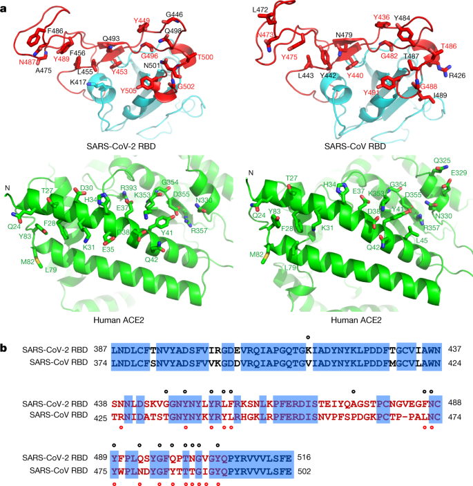
been carried out mainly on ACE2 and have confirmed that, like SARS-CoV, the S protein of SARS-CoV-2 uses ACE2 as a host surface receptor to enable the virus to enter and infect the cell.10,14–19 In addition to ACE2, CD147 protein has been identified as a co-receptor in host cells to enhance the ability of SARS-CoV-2 to enter human cells, and cause COVID-19 disease.11,14,16,17,20,21

In spite of these entry mechanisms, it was not clearly understood why the tissue tropism of SARS-CoV-2 varies from what is expected from virus–host cell interaction via ACE2 receptor, and why SARS-CoV-2 readily infects tissues other than the respiratory system, such as the brain, heart, and other tissues with no or low ACE2 expression. These raised the possibility that other host factors are required to facilitate virus–host cell interactions in cells with low ACE2 protein level.6,22 Thus, some studies were recently conducted to reveal these unclear mechanisms, and intriguingly, a major breakthrough study by Daly et al as well as a genetic based study by Cantuti-Castelvetri et al and Li and Buck identified a novel receptor called NRP1 that potentially answer the puzzling question what makes SARS-CoV-2 highly infectious and capable of rapidly spreading in human cells.6,22,23 They concluded that NRP1 may serve as an alternative or independent doorway for SARS-CoV-2 entry to and invasion of the human cells.

The Structure and Ligands of Neuropilin 1

NRP1, formerly known as A5 protein, is a 120–140 kDa non-tyrosine kinase multifunctional transmembrane heptameric protein with an ATWLPPR sequence consisting of 923 amino acids.1 It is encoded by the NRP1 gene located on the 10p11.22 human chromosome locus and shares 44% amino acid sequence identity with its homology, NRP2.3,4,24

NRP1 comprises a large N-terminus extracellular domain, a relatively very small plasma membrane spanning transmembrane domain, and a short cytoplasmic tail in the inner side of the cell membrane. The extracellular domain in turn comprised of three different subdomains, namely A, B, and C subdomains (Figure 1).1,3 The A or a1-a2 subdomains, also called semaphorin (SEMA) binding subdomains, are located on amino end and contains two complement binding motifs (CUB), namely the complement binding components (C1r and C1s), Uegf (urchin embryonic growth factor), and bone morphogenetic protein 1 (BMP1), whereas the B subdomain involving b1-b2 subdomains are found in the middle of the extracellular domain and are characteristics of clotting factor V and VIII, and discoidin proteins.25–27 This domain is also known as the vascular endothelial growth factors (VEGF) binding subdomain as it serves as a binding site for VEGF.1 The third domain, called C domain, has similarity with MAM (Meprin, A5/NRP1, protein tyrosine phosphatase μ and К) and contributes for homo- or hetero-dimerization of other receptors with the transmembrane domain, thereby affecting distinct downstream signaling cascades.3 The transmembrane domain is a single-pass protein consisting of a conserved GXXXG repeat crossing the cell membrane.27–29 A short domain of NRP1 that contains 43–44 amino acids but lacks tyrosine kinase activity is the cytosolic tail. This domain possesses a PDZ-binding motif that can interact with various proteins such RGS-GAIP-interacting protein (GIPC) and synectin that are essential for via VEGF receptor 2 (VEGFR2) signaling, arterial morphogenesis as well as in maintaining the structural integrity of transmembrane proteins.1,3,27

Figure 1 Schematic diagram of NRP1 Structure. NRP1 contains a large N-terminus extracellular domain comprising A (a1-a2), B (b1-b2), and C subdomains, a very small single-pass plasma membrane spanning TM domain, and a short cytoplasmic domain in the inner side of the cell membrane possessing PDZ-binding motif that can interact with various proteins. The a1/a2/b1 segment binds with SEMA3s, VEGFs, and other proteins, and the C domain involved in receptor dimerization with the TM domain. The b1 of NRP1 binds with S protein of SARS-CoV-2 and facilitate infection.

Abbreviations: NRP1, neuropilin 1; SEMA3s, class 3 semaphorin family; TM, transmembrane; VEGFs, vascular endothelial growth factors.

Although it lacks a direct cellular signaling role, NRP1 is most widely known for its crucial role as a multifunctional co-receptor by forming a complex with other membrane receptors to form holoreceptors.1 Numerous previous studies have shown that NRP1 is a membrane protein that serves as a surface receptor that can bind with wide varieties of protein families, including heparin-binding members of the VEGF family, class 3 members of the SEMA family (SEMA3s) such as SEMA3A, 3C, and 3F, transforming growth factor-β1 (TGF-β1).28,30,31 The SEMA3s are well known to bind to the a1/a2/b1 portion, whereas the VEGFs bind to b1/b2 segments of NRP1. In addition, NRP1 has recently been demonstrated to act as a receptor for extracellular microRNAs, fibroblast growth factor 2 (FGF-2), galectin-1, hepatocyte growth factor (HGF), plexin, β1 integrin, epidermal growth factor (EGF), platelet-derived growth factor (PDGF), hedgehog (Hh), and other growth factors, albeit their binding sites are not well characterized.32,33

Furthermore, a small portion of S protein is currently recognized to be complementary with the b1 domain of human NRP1 protein and allows the binding of SARS-CoV-2 to host cells and hence facilitate infection.6,22

The Role of Neuropilin 1 in Health and Disease

Many studies have pointed out that NRP1 is broadly distributed in the tissues of the human body, with a predominant expression in blood endothelial cells, vascular smooth muscle cells, mesenchymal stem cells, retinal vasculature, neurons, and epithelial cells lining the respiratory and gastrointestinal tracts.1,3,6,22,24 NRP1 has been established to play a myriad of physiological, pathological, and therapeutic roles. As indicated by the plethora of evidence, NRP1 has versatile functions in regulating a wide array of biological processes, such as axon guidance within the central and peripheral nervous systems, angiogenesis, vascular permeability, and cell survival, proliferation, differentiation, migration, and invasion.2,3,30,32

In recent years, NRP1 has also been shown to be expressed by various immune cells, such as macrophages, including alveolar, adipose tissue, bronchial, and vascular macrophages, dendritic cells, T-cells, particularly CD8 cells, regulatory T-cells, B-cells, and mast cells where it controls a multitude of functions, including development, migration, and recruitment, communication between different immune cells, as well as immune system regulation under normal physiological condition.3,4 NRP1 has also been detected in bone cells like osteoclasts and osteoblasts where it has an important role in regulating bone remodeling, for instance osteo-protection through its binding with SEMA3A.3,34,35

Besides, many other studies have improved the understanding of the roles of NRP1 in different pathological conditions, including cancer, immunological disorders, and bone diseases though the molecular mechanisms behind these functions are still to be elucidated.3,33,34 NRP1 is generally overexpressed in various clinical disorders, including malignancies, where it upregulates the oncogenic activities of malignant cells by enhancing cell survival and proliferation, and angiogenesis, as well as by contributing therapeutic resistance.1,3,33

Moreover, NRP1 has been investigated to have a potential role as a therapeutic target in various pathological disorders, for instance in cancer it serves as an antiangiogenic target as well as in cancer and autoimmune diseases it acts as a site for immunotherapies but it needs selective targeting of NRP1 under a particular clinical setting.3,33

Intriguingly, recent encroaching studies have unveiled an additional role of NRP1 in COVID-19 infection, which was found to be a cofactor and facilitator of SARS-CoV-2 entry and could pave the way to a new possible target of intervention for COVID-19.6,22 More recently, an NRP1 receptor was reported in one study as a potential new target for pain inhibitors to treat chronic pain.36 This discovery is based on the grounds of the pain-relieving activity of SARS-CoV-2, which is suggested to mediate the interactions of NRP1 with S-protein by preventing the normal binding of a protein called VEGF-A to NRP1 and blocking pain signals to give pain relief. However, before developing analgesics, more research is required on how NRP1 contributes to pain signaling.

Neuropilin 1 and COVID-19

Neuropilin 1 Mediated SARS-CoV-2 Entry Mechanism

According to recent studies, NRP1 is identified as a novel co-receptor as well as a potentiating factor for SARS-CoV-2 entry process by enhancing the interaction of the virus with ACE2 (Table 1).6,22,23,37 Daly et al suggested that it may act as an alternative doorway for SARS-CoV-2 to enter and infect human cells.22 Consistently, the study by Cantuti-Castelvetri et al using tissues from human autopsies revealed that NRP1 significantly potentiates SARS-CoV-2 infectivity.6 Furthermore, this study demonstrated that the NRP1 is expressed strongly in respiratory and olfactory epithelia, while the ACE2 is absent or expressed at low level and hence it may provide an independent gateway for viral entry and invasion of the host cells. Thus, the NRP1 may mediate SARS‑CoV‑2 entry into the brain via the olfactory bulb.6 In agreement with this, a study by Davies et al showed NRP1 is expression in the CNS, involving olfactory‑related regions such as the olfactory tubercles and para-olfactory gyri, suggesting the potential role of NRP1 as an additional mediator of SARS‑CoV‑2 infection implicated in the neurologic manifestations of COVID-19.37 Moreover, one review article has also suggested that the brainstem has a relatively high expression of ACE2 receptor, and possibly NRP1, that SARS-CoV-2 exploits for cell infection. Thus, the respiratory, cardiovascular, gastrointestinal, and neurological functions of the brainstem may be compromised indefinitely as a result of brainstem damage manifested with neurological symptoms even in mild cases of COVID-19 and may results in long-lasting consequences.38 Their abundant expression on epithelia exposed to the external environment as well as their multifaceted functions are the possible reasons that make NRP1 an ideal entry factor for SARS-CoV-2 as well as a critical contributing factor for multisystem involvement of SARS-CoV-2 infection.4,6,22

Table 1 A Summary Table on the Role of NRP1 in SARS-CoV-2 Infection

SARS-CoV-2 uses a small piece of S protein located on the outer surface of the virus to attach to a complementary region of NRP1 receptors on human cells and hence to penetrate the host cells (Figure 2).6 The cell entry phase of SARS-CoV-2 depends on priming of S protein by host cell proteases.39,40 At the S1/S2 boundary, there is a multi-basic sequence motif called RRAR containing arginine (R) and alanine (A) amino acids, with a sequence of Arg-Arg-Ala-Arg. The RRAR amino acid sequence is a unique feature of SARS-CoV-2 that provides a cleavage site for a host proprotein convertase (furin) and possibly form additional cell surface receptor binding sites and thus enhances pathogenicity by priming the fusion activity.39–41 This is confirmed by another study demonstrating that SARSCoV-2 virus with a natural deletion of the S1/S2 furin cleavage site is associated to attenuated pathogenicity in hamster models.42

Figure 2 The potential NRP1 mediated SARS-CoV-2 entry mechanism into human cells. The trimeric S protein of SARS-CoV-2 binds to host ACE2 via RBM of RBD. Furin mediated cleavage of S protein at S1/S2 site exposes the CendR motif of S1 and enables binding to the b1 subdomain of NRP1. Further processing of S protein by TMPRSS2 on the cell surface (early entry pathway) and CTSL in endolysosome (late entry pathway) exposes the FP and triggers membrane fusion, and the viral RNA get into the host cytoplasm. The genomic RNA undergoes replication and translation to form new SARS-CoV-2 virions after assembly in ERGIC and the new viruses finally released into the outside of the cell.

Abbreviations: ACE2, angiotensin converting enzyme 2; CendR, C-end rule; CT, cytoplasmic tail; CTD, C-terminal domain; CTSL, cathepsin L; FP, fusion peptide; HR, heptad repeat; NRP1, neuropilin 1; NTD, N-terminal domain; RBD, receptor binding domain; RBM, receptor binding motif; TM, transmembrane domain; TMPRSS2, TM serine protease 2; ERGIC, endoplasmic reticulum–golgi intermediate compartment.

The furin protease cuts the full-length S protein into S1 and S2 functional polypeptides and forms a multi-basic RRAR sequence on the carboxyl-terminal of S1 polypeptide. Studies based on x-ray crystallography and biochemical approaches have shown that the S1 C-end rule (CendR) motif is known to directly interact to b1 domain of NRP1 by electrostatic attraction and activate the cell surface receptors.4,6,22 However, even though the CendR peptide of S1 domain is a major recognition site for NRP1, it is not yet known which other segments of NRP1 and the S protein may interact. In addition, a comparable binding between the carboxy-terminal sequence of S1 subunit and NRP1 homolog, NRP2, was also demonstrated.22

A more recent study by Zhenlu and Matthias also indicated that NRP1 facilitate SARS-CoV-2 infection by stimulating the separation of S1 and S2 subunits.23 This study modeled the structures of NRP1 a2/b1/b2 binding to S protein of SARS-CoV-2 and showed NRP1 on binding with S protein trimer bound to an ACE2 dimer. The S1 binds more strongly to the host membrane in the presence of NRP1 and destabilizes the S1/S2 interface and hence increases the likelihood of the S2 subunit to be pulled out rather than S1 being stretched. Thus, NRP1 attachment may stimulate the easier dissociation of S2 from the S1 subunit that triggers membrane fusion and, thus, increase virus infectivity.23

Following the binding of ACE2 and NRP1 with S1 of SARS-CoV-2, further processing of S protein by transmembrane serine protease 2 (TMPRSS2) on the cell surface (early entry pathway) and cathepsin L(CTSL) in endolysosome (late entry pathway) occur. This exposes the fusion peptide (FP) that triggers membrane fusion, and the viral RNA get into the host cytoplasm. Then the genomic RNA undergoes replication and translation to form new virions after assembly in ERGIC, and the new viruses then are released into the outside of the cell.43,44 Previous studies showed that NRPs are known to mediate the internalization of CendR ligands through an endocytosis resembling micropinocytosis but it is unclear whether NRP1 allows attachment and receptor-mediated endocytosis in SARS-CoV-2 infected patients.6,22,45,46 Thus, further studies need to be done to clearly explain the role of NRP1 in the entry and infection mechanisms of SARS-CoV-2.

Swiss Model pandemisesta viruksesta sisältää paljon päivitettyä tietoa.europiliini 1 Sars-2 viruksen S1:n reseptorina .

 Sars-2 viruksen  neurotropismi oftalmiseen kudokseen on selitetty täten:  

Neuropiliinit NRP1 ja NRP2 toimivat myös  ACE2:n ohella  reseptoreina virukselle ja oftalmisessa kudoksessa ACE-2 pitoisuus on vähäinen ja neuropiliiniä taas esiintyy runsaammin. 

 Katson tietoa neuropiliinigeeneistä: 

 https://www.genecards.org/cgi-bin/carddisp.pl?gene=NRP1&keywords=NRP1

Entrez Gene Summary for NRP1 Gene

  • This gene encodes one of two neuropilins, which contain specific protein domains which allow them to participate in several different types of signaling pathways that control cell migration. Neuropilins contain a large N-terminal extracellular domain, made up of complement-binding, coagulation factor V/VIII, and meprin domains. These proteins also contains a short membrane-spanning domain and a small cytoplasmic domain. Neuropilins bind many ligands and various types of co-receptors; they affect cell survival, migration, and attraction. Some of the ligands and co-receptors bound by neuropilins are vascular endothelial growth factor (VEGF) and semaphorin family members. This protein has also been determined to act as a co-receptor for SARS-CoV-2 (which causes COVID-19) to infect host cells. [provided by RefSeq, Nov 2020]

GeneCards Summary for NRP1 Gene

NRP1 (Neuropilin 1) is a Protein Coding gene. Diseases associated with NRP1 include Covid-19 and Cerebral Arteriopathy, Autosomal Dominant, With Subcortical Infarcts And Leukoencephalopathy, Type 1. Among its related pathways are ERK Signaling and Development Slit-Robo signaling. Gene Ontology (GO) annotations related to this gene include heparin binding and growth factor binding. An important paralog of this gene is NRP2.


NRP2 on tunnewttu sytomegalovirusreseptorifunktiosta. NRP1 näyttää  olevan tas  Sars-2 virus reseptori.  

NRP2:

Entrez Gene Summary for NRP2 Gene

  • This gene encodes a member of the neuropilin family of receptor proteins. The encoded transmembrane protein binds to SEMA3C protein {sema domain, immunoglobulin domain (Ig), short basic domain, secreted, (semaphorin) 3C} and SEMA3F protein {sema domain, immunoglobulin domain (Ig), short basic domain, secreted, (semaphorin) 3F}, and interacts with vascular endothelial growth factor (VEGF). This protein may play a role in cardiovascular development, axon guidance, and tumorigenesis. Multiple transcript variants encoding distinct isoforms have been identified for this gene. [provided by RefSeq, Jul 2008]

GeneCards Summary for NRP2 Gene

NRP2 (Neuropilin 2) is a Protein Coding gene. Diseases associated with NRP2 include Wallerian Degeneration and Exudative Vitreoretinopathy 1. Among its related pathways are ERK Signaling and Signaling by GPCR. Gene Ontology (GO) annotations related to this gene include growth factor binding. An important paralog of this gene is NRP1.

UniProtKB/Swiss-Prot Summary for NRP2 Gene

  • High affinity receptor for semaphorins 3C, 3F, VEGF-165 and VEGF-145 isoforms of VEGF, and the PLGF-2 isoform of PGF.
  • (Microbial infection) Acts as a receptor for human cytomegalovirus pentamer-dependent entry in epithelial and endothelial cells.
 NRP1: https://www.dovepress.com/neuropilin-1-a-novel-entry-factor-for-sars-cov-2-infection-and-a-poten-peer-reviewed-fulltext-article-BTT

UniProtKB/Swiss-Prot Summary for NRP1 Gene

  • Cell-surface receptor involved in the development of the cardiovascular system, in angiogenesis, in the formation of certain neuronal circuits and in organogenesis outside the nervous system. Mediates the chemorepulsant activity of semaphorins (PubMed:9288753, PubMed:9529250, PubMed:10688880). Recognizes a C-end rule (CendR) motif R/KXXR/K on its ligands which causes cellular internalization and vascular leakage (PubMed:19805273). It binds to semaphorin 3A, the PLGF-2 isoform of PGF, the VEGF165 isoform of VEGFA and VEGFB (PubMed:9288753, PubMed:9529250, PubMed:10688880, PubMed:19805273). Coexpression with KDR results in increased VEGF165 binding to KDR as well as increased chemotaxis. Regulates VEGF-induced angiogenesis. Binding to VEGFA initiates a signaling pathway needed for motor neuron axon guidance and cell body migration, including for the caudal migration of facial motor neurons from rhombomere 4 to rhombomere 6 during embryonic development (By similarity). Regulates mitochondrial iron transport via interaction with ABCB8/MITOSUR (PubMed:30623799).
  • [Isoform 2]: Binds VEGF-165 and may inhibit its binding to cells (PubMed:10748121, PubMed:26503042). May induce apoptosis by sequestering VEGF-165 (PubMed:10748121). May bind as well various members of the semaphorin family. Its expression has an averse effect on blood vessel number and integrity.
  • (Microbial infection) Acts as a host factor for human coronavirus SARS-CoV-2 infection. Recognizes and binds to CendR motif RRAR on SARS-CoV-2 spike protein S1 which enhances SARS-CoV-2 infection.

lördag 26 juni 2021

SARS-2 pandemisen viruksen kiertävät variantit 23.6.2021

https://viralzone.expasy.org/9556 


Variants of Concern (VOC): Click to display variant sites (zoom in PDB window for a better view)

  • D614G
  • B.1.1.7 (Alpha)
  • B.1.351 (Beta)
  • P1 (Gamma)
  • B.1.617.2 (Delta)
  • B.1.427, B.1.429 (Epsilon)

Sars-2 viruksen S-proteiinin RBD ja RBM alueisiin ilmenneitä mutaatioita terapian etsinnän kannalta

(1) https://www.ncbi.nlm.nih.gov/pmc/articles/PMC7676316/ 

(2) https://www.nature.com/articles/s41392-021-00536-0/figures/1

Collectively, these results confirmed the capability of SARS-CoV-2 to escape from antibodies, especially the ACE2-competing antibodies, by acquiring resistance mutations in RBD. Such escape mutations can occur within the binding epitopes of antibodies, or regions away from antibody epitopes but may affect immunogenicity of RBD and render antibodies ineffective (Fig. 1f). Notably, the ACE2-competing antibodies constitute the majority of anti-RBD antibodies elicited by SARS-CoV-2 infection or vaccination. Therefore, our findings highlight the importance of continuously monitoring RBD natural mutations and evaluating their impact on antibody recognition of SARS-CoV-2, which may help guide the development and implementation of therapeutic antibodies and vaccines against SARS-CoV-2.

(3) Ilmoitus kaksoismutantti variaatiosta  https://www.biorxiv.org/content/10.1101/2020.03.15.991844v6

V367F + D614G  maaliskuussa 2020. 


(4) E484K mutaation merkityksestä artikkeli 

 https://www.thelancet.com/journals/lanmic/article/PIIS2666-5247(21)00068-9/fulltext

5) neljä päivää sitten tulut artikkeli Sars-2  mutaatioista ja varianteista:  https://viralzone.expasy.org/9556


Sars-2 viruksen S-proteiinin reseptoria sitova domeeni(RBD) ja reseptoriin kiinnittyvä motiivi(RBM)/ 2020 maaliskuu

 https://www.nature.com/articles/s41586-020-2180-5

Sitaatti 26.6.2021. Tässä artikkelissa vertaillaan SARS-2 ja Sars-1 virusten RBD ja RBM  kohtia.


Structure of the SARS-CoV-2 spike receptor-binding domain bound to the ACE2 receptor
Abstract

A new and highly pathogenic coronavirus (severe acute respiratory syndrome coronavirus-2, SARS-CoV-2) caused an outbreak in Wuhan city, Hubei province, China, starting from December 2019 that quickly spread nationwide and to other countries around the world1,2,3. Here, to better understand the initial step of infection at an atomic level, we determined the crystal structure of the receptor-binding domain (RBD) of the spike protein of SARS-CoV-2 bound to the cell receptor ACE2. The overall ACE2-binding mode of the SARS-CoV-2 RBD is nearly identical to that of the SARS-CoV RBD, which also uses ACE2 as the cell receptor4. Structural analysis identified residues in the SARS-CoV-2 RBD that are essential for ACE2 binding, the majority of which either are highly conserved or share similar side chain properties with those in the SARS-CoV RBD. Such similarity in structure and sequence strongly indicate convergent evolution between the SARS-CoV-2 and SARS-CoV RBDs for improved binding to ACE2, although SARS-CoV-2 does not cluster within SARS and SARS-related coronaviruses1,2,3,5. The epitopes of two SARS-CoV antibodies that target the RBD are also analysed for binding to the SARS-CoV-2 RBD, providing insights into the future identification of cross-reactive antibodies.

Main

The emergence of the highly pathogenic coronavirus SARS-CoV-2 in Wuhan and its rapid international spread has posed a serious global public-health emergency1,2,3. Similar to individuals who were infected by pathogenic SARS-CoV in 2003 and Middle East respiratory syndrome coronavirus (MERS-CoV) in 2012, patients infected by SARS-CoV-2 showed a range of symptoms including dry cough, fever, headache, dyspnoea and pneumonia with an estimated mortality rate ranging from 3 to 5%6,7,8. Since the initial outbreak in December of 2019, SARS-CoV-2 has spread throughout China and to more than 80 other countries and areas worldwide. As of 5 March 2020, 80,565 cases in China have been confirmed with the infection and 3,015 infected patients have died (https://www.who.int/emergencies/diseases/novel-coronavirus-2019/situation-reports/). As a result, the epicentre Wuhan and the neighbouring cities have been under lockdown to minimize the continued spread and the WHO (World Health Organization) has announced a Public Health Emergency of International Concern owing to the rapid and global dissemination of SARS-CoV-2.

Phylogenetic analyses of the coronavirus genomes have revealed that SARS-CoV-2 is a member of the Betacoronavirus genus, which includes SARS-CoV, MERS-CoV, bat SARS-related coronaviruses (SARSr-CoV), as well as others identified in humans and diverse animal species1,2,3,5. Bat coronavirus RaTG13 appears to be the closest relative of the SARS-CoV-2, sharing more than 93.1% sequence identity in the spike (S) gene. SARS-CoV and other SARSr-CoVs, however, are distinct from SARS-CoV-2 and share less than 80% sequence identity1.

Coronaviruses use the homotrimeric spike glycoprotein (comprising a S1 subunit and S2 subunit in each spike monomer) on the envelope to bind to their cellular receptors. Such binding triggers a cascade of events that leads to the fusion between cell and viral membranes for cell entry. Previous cryo-electron microscopy studies of the SARS-CoV spike protein and its interaction with the cell receptor ACE2 have shown that receptor binding induces the dissociation of the S1 with ACE2, prompting the S2 to transit from a metastable pre-fusion to a more-stable post-fusion state that is essential for membrane fusion9,10,11,12

Therefore, binding to the ACE2 receptor is a critical initial step for SARS-CoV to enter into target cells. Recent studies also highlighted the important role of ACE2 in mediating entry of SARS-CoV-21,13,14,15. HeLa cells expressing ACE2 are susceptible to SARS-CoV-2 infection whereas those without ACE2 are not1. In vitro binding measurements also showed that the SARS-CoV-2 RBD binds to ACE2 with an affinity in the low nanomolar range, indicating that the RBD is a key functional component within the S1 subunit that is responsible for binding of SARS-CoV-2 by ACE213,16.

The cryo-electron microscopy structure of the SARS-CoV-2 spike trimer has recently been reported in two independent studies13,17. However, inspection of one available spike structure revealed the incomplete modelling of the RBD, particularly for the receptor-binding motif (RBM) that interacts directly with ACE217. Computer modelling of the interaction between the SARS-CoV-2 RBD and ACE2 has identified some residues that are potentially involved in the interaction; however, the actual residues that mediate the interaction remained unclear18. Furthermore, despite detectable cross-reactive SARS-CoV-2-neutralizing activity of serum or plasma from patients who recovered from SARS-CoV infections15, no isolated SARS-CoV monoclonal antibodies are able to neutralize SARS-CoV-216,17. These findings highlight some of the intrinsic sequence and structure differences between the SARS-CoV and SARS-CoV-2 RBDs.

To elucidate the interaction between the SARS-CoV-2 RBD and ACE2 at a higher resolution, we determined the structure of the SARS-CoV-2 RBD–ACE2 complex using X-ray crystallography. This atomic-level structural information greatly improves our understanding of the interaction between SARS-CoV-2 and susceptible cells, provides a precise target for neutralizing antibodies, and assists the structure-based vaccine design that is urgently needed in the ongoing fight against SARS-CoV-2. Specifically, we expressed the SARS-CoV-2 RBD (residues Arg319–Phe541) (Fig. 1a, b) and the N-terminal peptidase domain of ACE2 (residues Ser19–Asp615) in Hi5 insect cells and purified them by Ni-NTA affinity purification and gel filtration (Extended Data Fig. 1). The structure of the complex was determined by molecular replacement using the SARS-CoV RBD and ACE2 structures as search models4, and refined to a resolution of 2.45 Å with final Rwork and Rfree factors of 19.6% and 23.7%, respectively (Extended Data Fig. 2 and Extended Data Table 1). The final model contains residues Thr333–Gly526 of the SARS-CoV-2 RBD, residues Ser19–Asp615 of the ACE2 N-terminal peptidase domain, one 

 ion, four N-acetyl-β-glucosaminide (NAG) glycans linked to ACE2 Asn90, Asn322 and Asn546 and to RBD Asn343, as well as 80 water molecules.

Fig. 1: Overall structure of SARS-CoV-2 RBD bound to ACE2.
figure1

a, Overall topology of the SARS-CoV-2 spike monomer. FP, fusion peptide; HR1, heptad repeat 1; HR2, heptad repeat 2; IC, intracellular domain; NTD, N-terminal domain; SD1, subdomain 1; SD2, subdomain 2; TM, transmembrane region. b, Sequence and secondary structures of SARS-CoV-2 RBD. The RBM sequence is shown in red. c, Overall structure of the SARS-CoV-2 RBD bound to ACE2. ACE2 is shown in green. The SARS-CoV-2 RBD core is shown in cyan and RBM in red. Disulfide bonds in the SARS-CoV-2 RBD are shown as sticks and indicated by arrows. The N-terminal helix of ACE2 responsible for binding is labelled.

The SARS-CoV-2 RBD has a twisted five-stranded antiparallel β sheet (β1, β2, β3, β4 and β7) with short connecting helices and loops that form the core (Fig. 1b, c). Between th

e β4 and β7 strands in the core, there is an extended insertion containing the short β5 and β6 strands, α4 and α5 helices and loops (Fig. 1b, c). This extended insertion is the RBM, which contains most of the contacting residues of SARS-CoV-2 that bind to ACE2. A total of nine cysteine residues are found in the RBD, eight of which form four pairs of disulfide bonds that are resolved in the final model. Among these four pairs, three are in the core (Cys336–Cys361, Cys379–Cys432 and Cys391–Cys525), which help to stabilize the β sheet structure (Fig. 1c); the remaining pair (Cys480–Cys488) connects the loops in the distal end of the RBM (Fig. 1c). The N-terminal peptidase domain of ACE2 has two lobes, forming the peptide substrate binding site between them. The extended RBM in the SARS-CoV-2 RBD contacts the bottom side of the small lobe of ACE2, with a concave outer surface in the RBM that accommodates the N-terminal helix of the ACE2 (Fig. 1c). The overall structure of the SARS-CoV-2 RBD is similar to that of the SARS-CoV RBD (Extended Data Fig. 3a), with a root mean square deviation (r.m.s.d.) of 1.2 Å for 174 aligned Cα atoms. Even in the RBM, which has more sequence variation, the overall structure is also highly similar (r.m.s.d. of 1.3 Å) to the SARS-CoV RBD, with only one obvious conformational change in the distal end (Extended Data Fig. 3a). The overall binding mode of the SARS-CoV-2 RBD to ACE2 is also nearly identical to that observed in the previously determined structure of the SARS-CoV RBD–ACE2 complex4 (Extended Data Fig. 3b).

The cradling of the N-terminal helix of ACE2 by the outer surface of the RBM results in a large buried surface of 1,687 Å2 (864 Å2 on the RBD and 823 Å2 on the ACE2) at the SARS-CoV-2 RBD–ACE2 interface. A highly similar buried surface of 1,699 Å2 contributed by SARS-CoV RBD (869 Å2) and ACE2 (830 Å2) is also observed at the SARS-CoV RBD–ACE2 interface. With a distance cut-off of 4 Å, a total of 17 residues of the RBD are in contact with 20 residues of ACE2 (Fig. 2a and Extended Data Table 2). Analysis of the interface between the SARS-CoV RBD and ACE2 revealed a total of 16 residues of the SARS-CoV RBD in contact with 20 residues of ACE2 (Fig. 2a and Extended Data Table 2). Among the 20 ACE2 residues that interact with the two different RBDs, 17 residues are shared between both interactions and most of the contacting residues are located at the N-terminal helix (Fig. 2a and Extended Data Table 2).

Fig. 2: The SARS-CoV-2 RBD–ACE2 and SARS-CoV RBD–ACE2 interfaces.
figure2

a, Contacting residues are shown as sticks at the SARS-CoV-2 RBD–ACE2 and SARS-CoV RBD–ACE2 interfaces. Positions in both RBDs that are involved in ACE2 binding are indicated by red labels. b, Sequence alignment of the SARS-CoV-2 and SARS-CoV RBDs. Contacting residues in the SARS-CoV-2 RBD are indicated by black dots; contacting residues in the SARS-CoV RBD are indicated by red dots.

To compare the ACE2-interacting residues on the SARS-CoV-2 and SARS-CoV RBDs, we used structure-guided sequence alignment and mapped them to their respective sequences (Fig. 2b). Among 14 shared amino acid positions used by both RBMs for the interaction with ACE2, 8 have the identical residues between the two RBDs, including Tyr449/Tyr436, Tyr453/Tyr440, Asn487/Asn473, Tyr489/Tyr475, Gly496/Gly482, Thr500/Thr486, Gly502/Gly488 and Tyr505/Tyr491 of SARS-CoV-2/SARS-CoV, respectively (Fig. 2b). Five positions have residues that have similar biochemical properties despite of having different side chains, including Leu455/Tyr442, Phe456/Leu443, Phe486/Leu472, Gln493/Asn479 and Asn501/Thr487 of SARS-CoV-2/SARS-CoV, respectively (Fig. 2b). The remaining position is at the Gln498/Tyr484 location (Fig. 2b), at which Gln498 of SARS-CoV-2 and Tyr484 of SARS-CoV both interact with Asp38, Tyr41, Gln42, Leu45 and Lys353 of ACE2. Among the six RBD positions with changed residues, SARS-CoV residues Tyr442, Leu472, Asn479 and Thr487 have previously been shown to be essential for binding ACE218. At the Leu455/Tyr442 position, Leu455 of SARS-CoV-2 and Tyr442 of SARS-CoV have similar interactions with Asp30, Lys31 and His34 of ACE2 (Fig. 3a). At the Phe486/Leu472 position, Phe486 of SARS-CoV-2 interacts with Gln24, Leu79, Met82 and Tyr83 of ACE2, whereas Leu472 of SARS-CoV has less interactions with Leu79 and Met82 of ACE2 (Fig. 3a). At the Gln493/Asn479 position, Gln493 of SARS-CoV-2 interacts with Lys31, His34 and Glu35 of ACE2 and forms a hydrogen bond with Glu35; Asn479 of SARS-CoV interacts with only His34 of ACE2 (Fig. 3a). At the Asn501/Thr487 position, both residues have similar interactions with Tyr41, Lys353, Gly354 and Asp355 of ACE2 (Fig. 3a). Asn501 of SARS-CoV-2 and Thr487 of SARS-CoV both form a hydrogen bond with Tyr41 of ACE2 (Fig. 3a). Outside the RBM, there is a unique ACE2-interacting residue (Lys417) in SARS-CoV-2, which forms salt-bridge interactions with Asp30 of ACE2 (Fig. 3b). This position is replaced by a valine in the SARS-CoV RBD that fails to participate in ACE2 binding (Figs. 2b, 3b). Furthermore, a comparison of the surface electrostatic potential also identified a positive charged patch on the SARS-CoV-2 RBD contributed by Lys417 that is absent on the SARS-CoV RBD (Fig. 3b). These subtly different ACE2 interactions may contribute to the difference in binding affinity of the SARS-CoV-2 and SARS-CoV to the ACE2 receptor (4.7 nM compared with 31 nM, respectively) (Extended Data Fig. 4).