Suurin osa, 80% toipunee SARS-kaltaisesta nykyinfektiosta- kertoo uutiset Kiinasta- kohtalaisen lievin oirein. Kuitenkin vakavia mainitaan olevan ainakin 20% erään tiedon mukaan. Kuolleisuuttatkin on, mutta sen osuutta ei vielä voi tarkentaa, ei kuitenkaan kovin katastrofaalinen ja yleensä vain niillä, joilla on muitakin tauteja . Päivittäinen kuolleisuus ilmoitetaan kymmenissä henkilöissä.
Vaikutaa siltä, että SARS infektio aiemmin , vaikka olisi ollut lievä, saattoi jättää jälkeensä keuhkoepiteelin korjaantumisen huononemaa monista molekulaarisesti selitettävistä syistä- ja minun mielestäni jälkihoito ja keuhkofunktion tarkistus joitain aikoja toipumisen akuutin jälkeenkin olisi aiheellinen. Ehkä jokin kortisonipitoinen astmalääkkeen tapainen jatkohoito saattaisi antaa epiteelille aikaa korjausmekanismeihin ja rauhoittaisi kiihtyneitä proteolyyttisiä järjestelmiä ja kollageenien muodostusta, ettei hengittävä pinta muutu dysfunktionaaliseksi.
https://books.google.se/books?id=ZEu4hd6YH2sC&pg=PA249&lpg=PA249&dq=MMPs+,+SARS-CoV&source=bl&ots=wDFcD592XO&sig=ACfU3U2ZNEkI6L3VMZQLgegpwRtVFIAYCQ&hl=sv&sa=X&ved=2ahUKEwj82uS-3aznAhVT4KYKHfrSAq4Q6AEwBnoECAcQAQ#v=onepage&q=MMPs%20%2C%20SARS-CoV&f=false
Hakusana (MMPS, SARS CoV) . Viite kirjasta, josta linkki on kovin pitkä litania kirjaimia
fredag 31 januari 2020
9b tumaan sukkulointi , Voisiko crm1 inhibitio estää sen sukkulointia? ?
crm1
(1) https://www.ncbi.nlm.nih.gov/pubmed/24631834
(1) https://www.ncbi.nlm.nih.gov/pubmed/24631834
Semin Cancer Biol. 2014 Aug;27:52-61. doi: 10.1016/j.semcancer.2014.03.002. Epub 2014 Mar 12.
Atomic basis of CRM1-cargo recognition, release and inhibition.
CRM1 or XPO1 is the major nuclear export receptor in the cell,
which controls the nuclear-cytoplasmic localization of many proteins
and RNAs. CRM1 is also a promising cancer drug target as the transport
receptor is overexpressed in many cancers where some of its cargos are
misregulated and mislocalized to the cytoplasm. Atomic level
understanding of CRM1 function has greatly facilitated recent drug
discovery and development of CRM1 inhibitors to target a variety of
malignancies. Numerous atomic resolution CRM1 structures are now
available, explaining how the exporter recognizes nuclear export signals
in its cargos, how RanGTP and cargo bind with positive cooperativity,
how RanBP1 causes release of export cargos in the cytoplasm and how
diverse inhibitors such as Leptomycin B and the new KPT-SINE compounds
block nuclear export. This review summarizes structure-function studies
that explain CRM1-cargo recognition, release and inhibition.DOI:10.1016/j.semcancer.2014.03.002
(3)
PLoS One. 2011;6(5):e19436. doi: 10.1371/journal.pone.0019436. Epub 2011 May 27.
SARS-CoV 9b protein diffuses into nucleus, undergoes active Crm1 mediated nucleocytoplasmic export and triggers apoptosis when retained in the nucleus.
Sharma K1, Åkerström S, Sharma AK, Chow VT, Teow S, Abrenica B, Booth SA, Booth TF, Mirazimi A, Lal SK.
9b is an accessory protein of the SARS-CoV. It is a small protein of 98 amino acids and its structure has been solved recently. 9b
is known to localize in the extra-nuclear region and has been
postulated to possess a nuclear export signal (NES), however the role of
NES in 9b functioning is not well understood.
In this report, we demonstrate that 9b in the absence of any nuclear localization signal (NLS) enters the nucleus by passive transport. Using various cell cycle inhibitors, we have shown that the nuclear entry of 9b is independent of the cell cycle. Further, we found that 9b interacts with the cellular protein Crm1 and gets exported out of the nucleus using an active NES. We have also revealed that this NES activity influences the half-life of 9b and affects host cell death. We found that an export signal deficient SARS-CoV 9b protein induces apoptosis in transiently transfected cells and showed elevated caspase-3 activity.
In this report, we demonstrate that 9b in the absence of any nuclear localization signal (NLS) enters the nucleus by passive transport. Using various cell cycle inhibitors, we have shown that the nuclear entry of 9b is independent of the cell cycle. Further, we found that 9b interacts with the cellular protein Crm1 and gets exported out of the nucleus using an active NES. We have also revealed that this NES activity influences the half-life of 9b and affects host cell death. We found that an export signal deficient SARS-CoV 9b protein induces apoptosis in transiently transfected cells and showed elevated caspase-3 activity.
Here, we showed that nuclear shuttling of 9b and its interaction with Crm1 are essential for the proper degradation of 9b and blocking the nuclear export of this protein induces apoptosis. This phenomenon may be critical in providing a novel role to the 9b accessory protein of SARS-CoV.
- PMID:
- 21637748
- PMCID:
- PMC3103500
- DOI:
- 10.1371/journal.pone.0019436
- [Indexed for MEDLINE]
torsdag 30 januari 2020
SARS CoV 9b , tumaan sukkuloiva Sars-lisäproteiini sammuttaa virustunnistussignalosomin
SARS-coronavirusopen reading frame-9bsuppresses innate immunity by targeting mitochondria and theMAVS/TRAF3/TRAF6 signalosome.
J Immunol. 2014 Sep 15;193(6):3080-9. doi: 10.4049/jimmunol.1303196. Epub 2014 Aug 18.
SARS-coronavirus open reading frame-9b suppresses innate immunity by targeting mitochondria and the MAVS/TRAF3/TRAF6 signalosome.
Coronaviruses
(CoV) have recently emerged as potentially serious pathogens that can
cause significant human morbidity and death. The severe acute
respiratory syndrome (SARS)-CoV was identified as the etiologic agent of the 2002-2003 international SARS outbreak. Yet, how SARS
evades innate immune responses to cause human disease remains poorly
understood.
In this study, we show that a protein encoded by SARS-CoV designated as open reading frame-9b (ORF-9b) localizes to mitochondria and causes mitochondrial elongation by triggering ubiquitination and proteasomal degradation of dynamin-like protein 1, a host protein involved in mitochondrial fission.
Also, acting on mitochondria, ORF-9b targets the mitochondrial-associated adaptor molecule MAVS signalosome by usurping PCBP2 and the HECT domain E3 ligase AIP4 to trigger the degradation of MAVS, TRAF3, and TRAF 6.
This severely limits host cell IFN responses.
Reducing either PCBP2 or AIP4 expression substantially reversed the ORF-9b-mediated reduction of MAVS and the suppression of antiviral transcriptional responses.
Finally, transient ORF-9b expression led to a strong induction of autophagy in cells. The induction of autophagy depended upon ATG5, a critical autophagy regulator, but the inhibition of MAVS signaling did not.
These results indicate that SARS-CoV ORF-9b manipulates host cell mitochondria and mitochondrial function to help evade host innate immunity.
This study has uncovered an important clue to the pathogenesis of SARS-CoV infection and illustrates the havoc that a small ORF can cause in cells.
- PMID:
- 25135833
- PMCID:
- PMC4179872
- DOI:
- 10.4049/jimmunol.1303196
- [Indexed for MEDLINE]
SARS-CoV lisäproteiineja on pystytty visualisoimaan 2015
Search results
Items: 16
- Kaksimolekulaarisesti fluoresoivalla komplementaatiomenetelmällä saatu näkyviin SARS-koronaviruksen lisäproteiineja. (2015)
Interactions among SARS-CoV accessory proteins revealed by bimolecular fluorescence complementation assay.
Acta Pharm Sin B. 2015 Sep;5(5):487-92. doi: 10.1016/j.apsb.2015.05.002. Epub 2015 Jun 6
Kong J1, Shi Y1, Wang Z1, Pan Y2. Abstract
- Suomennosta tiivistelmästä:
- SARS- koronaviruksen lisäproteiinit ( 3a, 3b, 6, 7a, 7b, 8a, 8b, 9b ja ORF14) ovat ennakoituja tuntemattomia proteiineja (PUP, predicted unknown proteins), joita geenit koodaavat ja joiden katsotaan olevan ainutlaatuisia jokaiselle SARS-CoV- virusgenomille, josta vaikeaa hengitystieoireyhtymää aiheutuu. Näillä proteiineilla on tärkeä osa erilaisissa biologisissa prosesseissa, joita välittää interaktiot näiden proteiinien ja niiden partnereitten kesken. Kuitenkin tiedetään hyvin vähän näistä lisäproteiinien keskuudessa vallitsevista interaktioista (vuorovaikutuksista).
- Tässä tutkimuksessa käytettiin bimolekulaarisesti keltaista fluoresoivaa proteiinien mittausmenetelmää (BiFC) (EYFP ) selviteltäessä näiden lisäproteiinien keskinäisiä vuorovaikutuksia. Voitiin tunnistaa 33 interaktiota 81:sta interaktiosta tällä kaksimolekulaarisella fluoresenssikomplementaatiomenetelmällä , mikä oli paljon enemmän kuin kaksihybridiseen hiivaan perustuvalla järjestelmällä (Y2H) saadut tulokset.
- Tämä on ensimmäinen raportti SARS-CoV- lisäproteiinien interaktioitten (vuorovaikutusten) visualisoimisesta, näkyviin saamisesta. Tämä viittaisi BiFC-systeemin yleiseen sovelluskelpoisuuteen proteiini-proteiini-interaktioitten todentamisessa.
The accessory proteins (3a, 3b, 6, 7a, 7b, 8a, 8b, 9b and ORF14), predicted unknown proteins (PUPs) encoded by the genes, are considered to be unique to the severe acute respiratory syndrome coronavirus (SARS-CoV) genome. These proteins play important roles in various biological processes mediated by interactions with their partners. However, very little is known about the interactions among these accessory proteins. Here, a EYFP (enhanced yellow fluorescent protein) bimolecular fluorescence complementation (BiFC) assay was used to detect the interactions among accessory proteins. 33 out of 81 interactions were identified by BiFC, much more than that identified by the yeast two-hybrid (Y2H) system. This is the first report describing direct visualization of interactions among accessory proteins of SARS-CoV. These findings attest to the general applicability of the BiFC system for the verification of protein-protein interactions.
Avainsanoja, KEYWORDS:
Aktivaatiodomeeni, AD, activation domain;Lisäproteiineja, (Apuproteiineja), Accessory proteins;
Sitova domeeni, BD, binding domain;
kaksimolekulaarinen fluoresenssikomplementaatiomenetelmä , BiFC, bimolecular fluorescence complementaatio, Bimolecular fluorescence complementation assay;https://de.wikipedia.org/wiki/Bimolekulare_Fluoreszenzkomplementation https://www.fpbase.org/protein/eyfp/
proteiinikompleksin immunosaostus , Co-IP, Co-immunoprecipitation; Protein complex immunoprecipitation (Co-IP)
vaippa, E, envelope; (Vaippaproteiinin merkki)
Vaippa Joillain viruksilla nukleokapsidia ympäröi lipideistä koostuva kaksoiskerroksinen kalvorakenne, vaippa, joka on peräisin isäntäsolusta. Vaippaan on uponneena viruksen koodaamia proteiineja, yleensä glykoproteiineja, joilla on tehtävä isäntäsolun tunnistamisessa ja infektion aloitamisessa.(Ruotsiksi Lipidhölje)
vahvistetusti keltaista fluoresoiva proteiini, EYFP, enhanced yellow fluorescent protein;
kalvo, M, membrane;kalvoproteiinin (M) merkki
nukleokapsidi, N, nucleocapsid;nukleokapsidiproteiinin merkki (N)
tumaan paikallistava signaali, NLS, nuclear localization signal;
aukinaisia luettavissa olevia lukuraameja, avoimia lukukehiöitä, ORFs, open reading frames;
polymeraasiketjureaktio, PCR, polymerase chain reaction;
proteiinien keskinäisiä vuorovaikutuksia, PPIs, protein-protein interactions;
ennustettavissa olevia tuntemattomia proteiineja, PUPs, predicted unknown proteins;
piikki, S, spike;
vaikeaa hengitystieoireyhtymää aiheuttava koronavirus SARS-CoV; SARS-CoV, severe acute respiratory syndrome coronavirus;
kaksihybridinen hiivamenetelmä Y2H; Y2H, yeast two-hybrid;
aminohappo, aa, amino acids
- PMID:
- 26579480
- PMCID:
- PMC4629423
- DOI:
- 10.1016/j.apsb.2015.05.002
SARS-CoV lisäproteiineista artikkeleita edelleen 9b syntyy alternatiivilla lukuraamilla orf9a
Similar articles
Select item 254100512.
Acquisition of new protein domains by coronaviruses: analysis of overlapping genes coding for proteins N and 9b in SARS coronavirus.
Shukla A, Hilgenfeld R.
Virus Genes. 2015 Feb;50(1):29-38. doi: 10.1007/s11262-014-1139-8. Epub 2014 Nov 20.
Acquisition of new protein domains by coronaviruses: analysis of overlapping genes coding for proteins N and 9b in SARS coronavirus. Shukla A1, Hilgenfeld R. Abstract
- Tavallisesti- kun virukset hankkivat uusia kykyjä genomipotentiaaliinsa, ne tekevät horisontaalisia geenin siirtoja tai kaksikertaistavat geeniä. Mutta eräs epätavallisempi keino on käyttää täysin tai osittain toisiansa kattavia avoimia lukukehyksiä (ORF,open reading frames).
- SARS.koronavirus on tälla keinolla sellaisen kokonaan uuden proteinituotteen hankinnan, että se on saanut uuden nimenkin 9b ja siinä SARS CoV on johtanut aloituskodonin vaihtoehtoisen lukukehyksen alueelle. Tämä geeni kattaa täydellisesti nukleokapsidiproteiinin (N) geenin avoimen lukukehyksen (orf9a ).
- Tutkijoiden löytö viittaa siiheen, että orf9b antaa "sellaiset soinnut, jotkä eivät ole saman säveltä muun SARS- CoV:n sinfoniassa", vaan siinä on jotakin aivan erilaista tässä viruksen työtiimissä. He analysoivat orf9b:n evoluutiota yhdessä orf9a kanssa käyttämällä sekvenssitietueita betakoronaviruslinjasta b ja havaitsivat, että orf9b, joka koodaa päältäkirjoittuvaa proteiinia, kehittyi suurelta osin orf9a kohdan päältä kirjoitetusta proteiinista itsenäisesti Sitten tutkijat selvittivät edelleen näiden genomisekvenssien proteiinituotteet niiden rakenteellisen joustavuuden kannalta ja havaitsivat, että tämän äskettäin hankitun geenialueen kattavan proteiinituotteen ei tarvitse välttämättä olla edes sisäisesti järjestäytyneenä, vaikka aiemmin on niin ajateltu. Nämä löydöt osaltaan auttavat luonnehtimaan äskettäin hankittujen geenien sekvenssiominaisuuksia käyttäällä hyödyksi päällekkäisiä avoimia lukukehyksiä.
Acquisition
of new proteins by viruses usually occurs through horizontal gene
transfer or through gene duplication, but another, less common mechanism
is the usage of completely or partially overlapping reading frames (orf).
A case of acquisition of a completely new protein through introduction of a start codon in an alternative reading frame is the protein encoded by open reading frame (orf) 9b of SARS coronavirus. This gene completely overlaps with the nucleocapsid (N) gene (orf9a).
Our findings indicate that the orf9b gene features a discordant codon-usage pattern. We analyzed the evolution of orf9b in concert with orf9a using sequence data of betacoronavirus-lineage b and found that orf9b, which encodes the overprinting protein, evolved largely independent of the overprinted orf9a. We also examined the protein products of these genomic sequences for their structural flexibility and found that it is not necessary for a newly acquired, overlapping protein product to be intrinsically disordered, in contrast to earlier suggestions. Our findings contribute to characterizing sequence properties of newly acquired genes making use of overlapping reading frames.
- PMID:
- 25410051
- DOI:
- 10.1007/s11262-014-1139-8
- [Indexed for MEDLINE]
Englantilaiset uutiset Kiinasta 01.38, 30.1. 2020
https://www.chinadaily.com.cn/index.html?fbclid=IwAR3kzQXutsYvT9GP4woKdhWHvx6wcnNG7y_WcCjHHevXs-y
The number of confirmed cases of the novel coronavirus on the Chinese mainland has exceeded the total number of SARS cases recorded during that outbreak 17 years ago. But there is no need to panic, experts said on Wednesday, even though more cases are expected over the next few days.
The number of confirmed cases of the new coronavirus reached 5,974 on Tuesday — an increase of 1,459 over the day before, including 132 deaths, since the outbreak was first reported late December, according to the National Health Commission on Wednesday.
In addition, the number of suspected cases rose to 9,239.
In Hubei province, the epicenter of the outbreak, 840 new cases were reported on Tuesday, bringing the total number of confirmed cases there to 3,554.
A suspected case was reported on Tuesday in the Tibet autonomous region, which, if confirmed, would mean that every province, autonomous region and municipality on the mainland has acquired the novel coronavirus.
Wednesday's data mean the new virus has exceeded the spread of SARS in its first month.
With SARS — severe acute respiratory syndrome — a total of 5,327 severe cases were reported on the Chinese mainland between the end of 2002 and Aug 16, 2003, including 349 deaths, according to what was then the Ministry of Health.
In addition, cases of infected foreigners on the mainland were first reported in South China's Guangdong province.
Three foreigners — a Pakistani and two Australians, who all had been in Wuhan recently — have been diagnosed with the coronavirus in Guangdong province as of Wednesday.
Guangzhou released on Wednesday a public letter and details of multilingual service hotlines for foreigners to get help.
Wuhan and Tianjin also have provided foreigners with timely consultations and assistance on epidemic prevention and control by opening a 24-hour hotline service.
Zhong Nanshan, a prominent expert in respiratory diseases and a member of the Chinese Academy of Engineering, said the recent novel coronavirus outbreak may hit its peak in a week or 10 days. The outbreak will not last as long as the SARS outbreak — more than five months — in part because of strong measures to contain the outbreak adopted by the central government, he said.
There are still no effective drugs to combat the virus, but researchers and medical staff have been working on several methods, and life-support technologies have improved greatly since SARS, so the death rate will be less, he told Xinhua News Agency.
So far, there has been no official statement about when the epidemic will peak or how long it will last. Gauden Galea, the World Health Organization's representative in China, said in an earlier interview with China Daily that the WHO is organizing a number of researchers to model the case numbers, but no conclusion has been reached yet.
Zeng Guang, chief epidemiologist at the Chinese Center for Disease Control and Prevention, said that compared with SARS, which involved many critical cases, the recent coronavirus outbreak is less severe. People in a large number of confirmed cases showed mild symptoms, according to a report in Health Times on Wednesday.
However, the new virus is more difficult to control and prevent than SARS. It can jump between humans during incubation, which lasts up to 14 days, he said.
With the lockdown of Wuhan, Hubei province, the presumed source of the outbreak, the number of cases exported from the city will gradually be reduced, and the rise in cases in other parts of China will likewise slow, Zeng said.
Meanwhile, warming weather will also restrain the spread of respiratory diseases and contribute to control and prevention, he said.
The WHO said in a statement on Tuesday that studies so far indicate that most cases of the virus reported to date have been milder, with about 20 percent of all confirmed cases experiencing severe illness.
China's National Health Commission will continue to collaborate with the WHO to contain the outbreak, including studying the severity and transmissibility of the virus, it said. In addition, China will share biological material with the WHO to contribute to the development of vaccines.
The WHO will also send international experts to visit China as soon as possible to work with domestic experts to increase understanding of the outbreak and guide global response efforts, it said.
The statement came after a meeting between President Xi Jinping and Tedros Adhanom Ghebreyesus, director-general of the WHO, in Beijing on Tuesday.
"We appreciate the seriousness with which China is taking this outbreak, especially the commitment from top leadership and the transparency they have demonstrated, including sharing data and the genetic sequence of the virus," the statement said. "The WHO will keep working side-by-side with China and all other countries to protect health and keep people safe."
"Both the WHO and China noted that the number of cases being reported, including those outside China, is deeply concerning," the statement said. "Better understanding of the transmissibility and severity of the virus is urgently required to guide other countries on appropriate response measures."
Walter Ian Lipkin, a professor of epidemiology and director of the Center for Infection and Immunity at Columbia's Mailman School of Public Health who is known as a leading "virus hunter", is headed to the epidemic-stricken area in China to assist with efforts to contain the spread of the novel coronavirus, according to Columbia Global Centers in Beijing on Tuesday.
Based on the evidence so far, Lipkin said on Tuesday in an article updated on Columbia University's website that the novel coronavirus is not expected to spread to the same extent as SARS, which reached 33 countries.
Sara Åkeström et al. työryhmä SARS-coV viruksen lisäproteiineja selvittämässä . 9b selvitetty
Lainakirja jonka viimeksi lainasin on sara Åkerströmin väitöstyö vuodelta 2008:
Sars Coronavirus The role of accessory proteins and nitric oxide in the replication cycle.
Tässä kun olen katsonut viimeksi 3a -lisäproteiinin kykyjä, pohdin sitä materiaalia, mikä viruksen "hiljaisen" ( kliinissti oirettoman inkubaatioajan kuluessa ehtii virrata tuman sisätilaankin ja etsin tämän materiaalin nimeä. Löysin artikkelin lisäproteiinista 9b ja työssä (2011 julkaisun nimiluettelossa) näkyi olevan mukana Sara Åkerström. Siteeraan tämän artikkelin tähän kohtaan:
Research Article (Nimi suomennettuna) "SARS-CoV 9b proteiini diffundoituu tumaan ja käy läpi aktiivin Crm1-välitteisen nukleosytoplasmisen uloskuljetuksen ja triggeröi ohjelmoidun solukuoleman eli apoptoosin , jos joutuu pysyttelemään tumassa".
Nyt on kello jo yli puolen yön, muta koska tuo 9b proteiinikin on sellainen kellonaikoja noudattava, niin jatkan tätä kirjoittelua. Sain kahlattua päälisin puolin tuon pienen lisäproteiinin tarinaa. Sitä ilmenee Sars-Co Viruksen infektoimassa solussa ja sille on osoitettavissa vasta-aineita. Sen rakenteessa piilee NES, tumasta ulos päin kohdentava osoitelappu, mutta ei tumaan sisään kohdentavaa lappua (NLS). Sillä ei varsinaisesti ole suuria struktuuriproteiinin tehtäviä kuten S, M, N ja E- proteiineilla, mutta se on SARS-CoV- rakenteessa kuin kissan häntä mukana ja suorittaa ikäänkuin turhan näköistä sukkulointia tuman ja sytoplasman välillä. Se menee passiivisiseti tumaan ilmeisesti riippuen konsentraatiostaankin(?) ja siellä sitten ilmenee sen NES ja se liittyy isäntäkehon lojaaleihin tumasta uloskuljettaviin proteiineihin ja tulee siten ulos tumastakuin "omalla avaimella , vaikka on mennyt sinne jotenkin salaa ilman näennäistä avainta, siis diffuusiolla.
12 tuntia infektoitumisesta sitä esiintyy sytoplasmssa havaittavia määriä. 24 tunnin kulutua ilmenee myös tumassa lievää värjytymistä. Solussa esiintyminen saavuttaa piikkinsä 36 tunnin kuluttua, jolloin tumassa on lukuisia värjäytyneitä fokuksia ja sytoplasmassa laajalla alueella inkluusioita ja 48 tunnin kuluttua on tapahtunut tumassa jonkin verran laskua pitoisuuksissa. Sitten tämä sukkkulointi jatkuu jatkumistaan. Tumasta uloskuljettaminen merkitsee proteiinien kuljetusta degradaatiojärjestelmään, ubikitinaatioon ja proteosomaaliseen hajoitukseen. Jos nyt siten 9b menee tähän ubikitinoitumiseen, tietysti Sarsin deubikitinaasi käsittelee sen, sillä sars pystyy sekä ubikitinoimaan että deubikitinoimaan systeminsä jäseniä tarpeen mukaan ajallaan. Jos 9b määrä on matala, olisi loogista että nsp3 deubikitinoisen ja se jatkaa tuota passiivia tumaan menoa. samalla on ubikitinaatiojärjestelmän polttopisteet paljastuneet Sars-virukselle ja se voi vähä vähältää toimitaa solunsisäisen antivirusjärjestelmän tehottomaksi tekemisen tämän luotaimen avulla. Kun sitten on lopulta virussukupolvi valmiina tuma voidaan vielä hajoitaa myös alkuaineikseen 9b:n avulla. Se kertyessään tumaan asettaa solun apoptoitumaan ja kaspaasit hajoittavat proteiinit, sillä sarsviruksen aminohappojen tarve tarve on todella suuri. Tuleeko tämä kissanhännän pää sitten pakattua virioniin se on kyseenalaista, koska se kuitenkin ilmenee uudelleen genomista tietä solussa joka infektoituu. Sitä ei tarvitse pakata mukaan proteiinimuodossa. Loogista. Sars infektio ei vaadi aktiivia solusykliä, solu voi olla Go vaiheessa ja tämä toimii silti hyvin. Olisi mahdollista myös meningiitti ja muut hermokudoksen tulehdukset myös. Tokko keuhkoalveolirakenteenkaan soluilla mikään suuri tumasykli on. Minusta virus on erittäin tuhoisa. Voisi sanoa Biologisen aseen luokkaa. Täytyy tarkistaa löytyykö aivotulehduksista tietoa. Ja täytyy vain toivoa että uusi 2019-nCoV ei ole hankkinut lisäkykyjä siltä kapasiteetilta neurotrooppisilta kannoilta. Täytyy seurata uutisia, mitä tiedemiehet kertovat uudesta viruksesta.
Sars Coronavirus The role of accessory proteins and nitric oxide in the replication cycle.
Tässä kun olen katsonut viimeksi 3a -lisäproteiinin kykyjä, pohdin sitä materiaalia, mikä viruksen "hiljaisen" ( kliinissti oirettoman inkubaatioajan kuluessa ehtii virrata tuman sisätilaankin ja etsin tämän materiaalin nimeä. Löysin artikkelin lisäproteiinista 9b ja työssä (2011 julkaisun nimiluettelossa) näkyi olevan mukana Sara Åkerström. Siteeraan tämän artikkelin tähän kohtaan:
Research Article (Nimi suomennettuna) "SARS-CoV 9b proteiini diffundoituu tumaan ja käy läpi aktiivin Crm1-välitteisen nukleosytoplasmisen uloskuljetuksen ja triggeröi ohjelmoidun solukuoleman eli apoptoosin , jos joutuu pysyttelemään tumassa".
- Published: May 27, 2011
- https://doi.org/10.1371/journal.pone.0019436
12 tuntia infektoitumisesta sitä esiintyy sytoplasmssa havaittavia määriä. 24 tunnin kulutua ilmenee myös tumassa lievää värjytymistä. Solussa esiintyminen saavuttaa piikkinsä 36 tunnin kuluttua, jolloin tumassa on lukuisia värjäytyneitä fokuksia ja sytoplasmassa laajalla alueella inkluusioita ja 48 tunnin kuluttua on tapahtunut tumassa jonkin verran laskua pitoisuuksissa. Sitten tämä sukkkulointi jatkuu jatkumistaan. Tumasta uloskuljettaminen merkitsee proteiinien kuljetusta degradaatiojärjestelmään, ubikitinaatioon ja proteosomaaliseen hajoitukseen. Jos nyt siten 9b menee tähän ubikitinoitumiseen, tietysti Sarsin deubikitinaasi käsittelee sen, sillä sars pystyy sekä ubikitinoimaan että deubikitinoimaan systeminsä jäseniä tarpeen mukaan ajallaan. Jos 9b määrä on matala, olisi loogista että nsp3 deubikitinoisen ja se jatkaa tuota passiivia tumaan menoa. samalla on ubikitinaatiojärjestelmän polttopisteet paljastuneet Sars-virukselle ja se voi vähä vähältää toimitaa solunsisäisen antivirusjärjestelmän tehottomaksi tekemisen tämän luotaimen avulla. Kun sitten on lopulta virussukupolvi valmiina tuma voidaan vielä hajoitaa myös alkuaineikseen 9b:n avulla. Se kertyessään tumaan asettaa solun apoptoitumaan ja kaspaasit hajoittavat proteiinit, sillä sarsviruksen aminohappojen tarve tarve on todella suuri. Tuleeko tämä kissanhännän pää sitten pakattua virioniin se on kyseenalaista, koska se kuitenkin ilmenee uudelleen genomista tietä solussa joka infektoituu. Sitä ei tarvitse pakata mukaan proteiinimuodossa. Loogista. Sars infektio ei vaadi aktiivia solusykliä, solu voi olla Go vaiheessa ja tämä toimii silti hyvin. Olisi mahdollista myös meningiitti ja muut hermokudoksen tulehdukset myös. Tokko keuhkoalveolirakenteenkaan soluilla mikään suuri tumasykli on. Minusta virus on erittäin tuhoisa. Voisi sanoa Biologisen aseen luokkaa. Täytyy tarkistaa löytyykö aivotulehduksista tietoa. Ja täytyy vain toivoa että uusi 2019-nCoV ei ole hankkinut lisäkykyjä siltä kapasiteetilta neurotrooppisilta kannoilta. Täytyy seurata uutisia, mitä tiedemiehet kertovat uudesta viruksesta.
- J Neuropathol Exp Neurol. 1999 Dec;58(12):1197-206.
- Nidovirus infections: experimental model systems of human neurologic diseases.
- Lavi E1, Schwartz T, Jin YP, Fu L.Abstract
- The presence of terminally differentiated slow- and non-dividing cells in the central nervous system (CNS) provides a safe harbor for viral persistence and latency and constitutes a unique immunologic environment for viral infections. Studies of experimental model systems of viral infections of the CNS provide insight into mechanisms of viral persistence and immune-mediated pathology. Nidoviruses are comprised of 2 families of viruses, coronaviruses and arteriviruses, and are common pathogens of humans and a variety of animal species. Both families of viruses contain neurotropic strains that produce experimental neurologic diseases in rodents. These include acute meningitis and encephalitis; acute poliomyelitis; and chronic inflammatory, immune-mediated, demyelination. Coronavirus-induced demyelinating disease mimics many of the pathologic features of Multiple Sclerosis (MS).
- PMID:
- 10604745
- DOI:
- 10.1097/00005072-199912000-00001
- [Indexed for MEDLINE]
onsdag 29 januari 2020
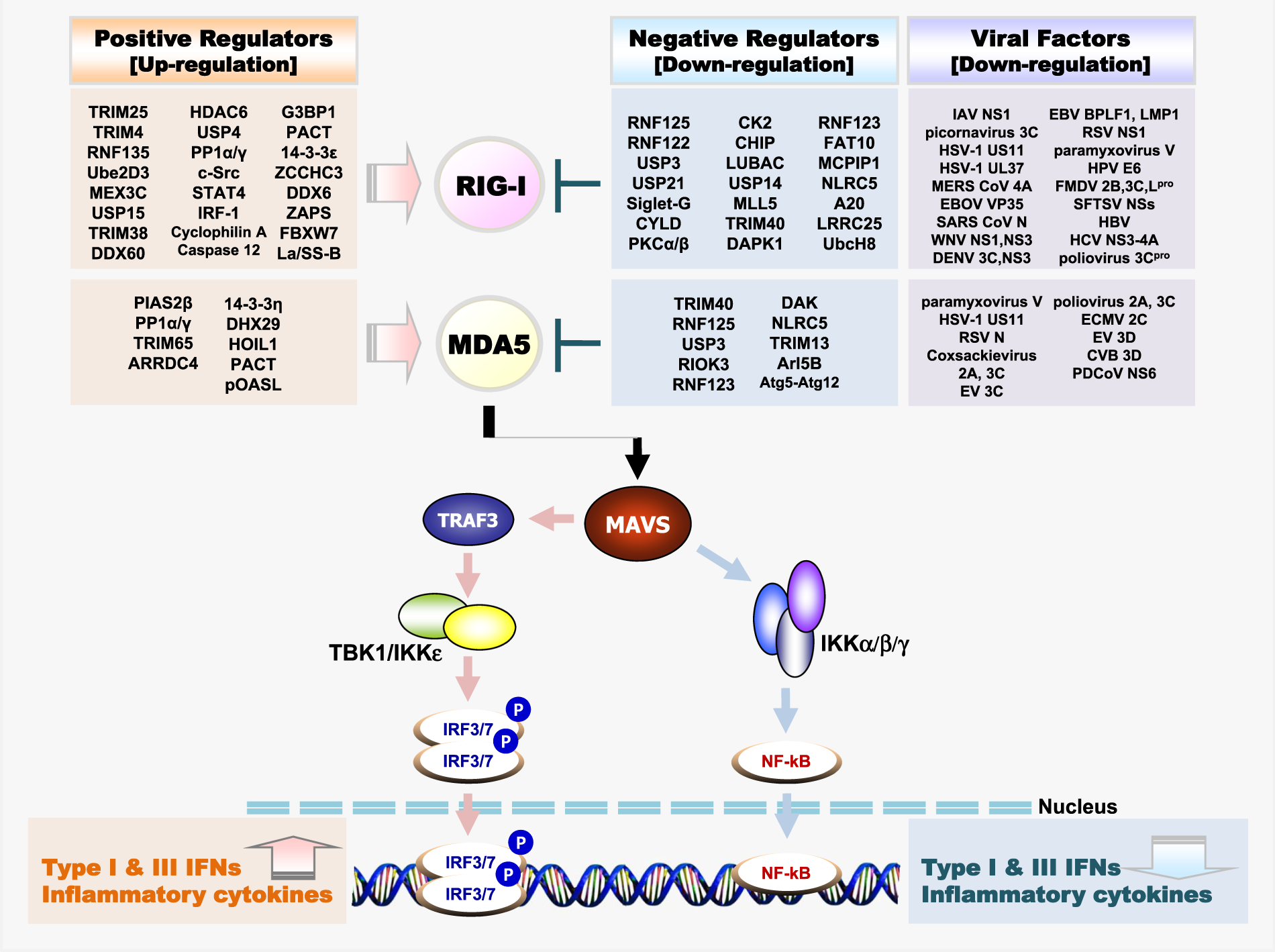
TRIM- proteiinit ja RIG1, MDA5 , Solunsisäistä vieraan RNA:n tunnistusjärjestelmää
https://media.springernature.com/full/springer-static/image/art%3A10.1038%2Fs12276-019-0299-y/MediaObjects/12276_2019_299_Fig1_HTML.png
Tämän järjestelmän SARS-CoV pystyy kiertämään monipuolisen tehokkaalla evaasiolla.
Tämän järjestelmän SARS-CoV pystyy kiertämään monipuolisen tehokkaalla evaasiolla.
NOD ja NOD-Like reseptoreita, inflammasomin aktivoituminen ja interleukiinien tuotanto
https://www.bocsci.com/upload/image/NOD-like-signal-pathway-3.JPG
Tässä on yksi kuva NOD-reseptoriperheen NLRP3:n inflammasomin aktivoitumsesta.
Tässä on yksi kuva NOD-reseptoriperheen NLRP3:n inflammasomin aktivoitumsesta.
SARS-CoV 3a-lisäproteiini aktivoi viroporilnillaan NOD- proteiineihin kuuluvan reseptorin , inflammasomin ja interleukiineja IL
29.1.
2020
SARS-CoV lisäproteiini 3a omaa myös viaporiini kanavan kuten E ja voi aktivoida NOD-kaltaisia reseptoreita ja tätä tietä inflammasomin ja herättää interleukiinieritystä, sytokiineja. Arvellaan että tämä myöhäisfunktio toimii viruksen E- proteiinifunktion vaimentajana siinä vaiheessa kun virionit jo valmistuvat ja ovat lähdössä pois solusta, jolloin on eduksi saada funktion vaimennuksia. E-proteiini taas on saanut aikansa stabiiliutta itselleen ubikitinaastiosta, jonka virus itse tekee nsp3- tietä ja samalla deubikitinoi isäntäsolun järjstelmiä ja suppressoi täysin IFN- järjestelmän ja ISG15 välitteiset vaikutukset.
Nsp3:n DUB- ubikitiiniligaasi- duaalifunktiossa on jotain samanlaista kuten OTU ryhmässäkin, jossa löytyy duaaleja DUBeja ( siis saman proteiinin toisessa päässä on deubikitinointikykyä ja toisessa ubikitinointikykyä. Lisäksi Nsp3 deisgyloi. ISG15 on rakenteeltaan kuin di-ubikitiini: kaksi ubikitiiniketjua peräkkäin.
Tietysti E vaimenee, jos virus itse deubikitinoisi myöhäisvaiheens, mutta kapea-alainen vaimentaminen jollain spesiaalityöntekijä, lisäproteiinilla, on loogista. ja E-proteiini saa hoitaa virionit perille asti. Loppuvaiheessa (histopatol) keuhkokudoksessa nähdään näitä neutrofiileja, makrofageja, ja mitä nyt inflammasomiaktivaatiosta voi tulla.
https://www.ncbi.nlm.nih.gov/pubmed/?term=Coronavirus+Inflammasome%2C+NOD
Front Microbiol. 2019 Jan 29;10:50. doi: 10.3389/fmicb.2019.00050. eCollection 2019.
Severe Acute Respiratory Syndrome Coronavirus Viroporin 3a Activates the NLRP3 Inflammasome.
Suomennosta tiivistelmästä:
NODin kaltaiset reseptoriperhe, pyriinidomeenin3 sisältävä NLRP3
säätelee interleukiinien IL1beta ja IL-18 erityistä.
Nämä interleukiinit ovat proinflammatorisia sytokiineja.
Tutkijaryhmä on aiemmin osoittanut, että influenssaviruksen membeaaniproteiini M2 tai enkefalomyokardiittiviruksen (EMCV) 2b-proteiini stimuloivat interleukiinin IL-1beta eritystä, kun inflammasomi NLRP3 aktivoitui Mutta SARS-koronaviruksen mekanismia, jolla se saa aikaan NLRP3:n aktivoitumisen , ei ole aiemmin tunnettu.
Tässä työssään tutkijat pystyvät antamaan suoraa näyttöä siitä, että SARS-CoV 3a proteiini aktivoi inflammasomin NLRP3 sellaisissa makrofageissa, joissa oli LPS primeeraus. SARS-CoV 3a oli riittävä tekijä aiheuttamaan inflamamsomin NLRP3 aktivaation. Tässä 3a-pvälitteisessä- interleukiinin IL-1beta erityksessä oli 3a-proteiinin jonikanavan aktiivisuus essentielli, välttämätön seikka. Infektoitumattomissa soluissa tai lentiviruksella infektoituneissa soluissa, joissa oli jonikanava-aktiivisuudelta vajeinen 3a-proteiini, nämä inflammasomit NLRP3 sijaitsivat tasaisesti jakaantuneena sytoplasmaan . Mutta jos infektoituneissa lentiviruksissa oli kunnolla toimivan jonikanavan omaava 3a-proteiini, NLRP3-inflammasomit järjestäytyivät uudelleen asettuen solujen perinukleaariseen tilaan, siis tuman lähistöön. NLRP3- Inflammasomien SARS-CoV 3a- aktivaatiossa oli tärkeänä tekijänä Kaliumjonien (K+) ulosvirtaus ja ROS ( reaktiiviset happilajit). Nämä tulokset valaisevat viroporiinien tärkeyttä transmembraanisina (kalvon läpi johtavina) aukkoa muodostavina virusproteiineina viruksen alkuunsaamissa NLRP3-inflammasomien aktivoimisessa.
- Nod-like receptor family, pyrin domain-containing 3 (NLRP3) regulates the secretion of proinflammatory cytokines interleukin 1 beta (IL-1β) and IL-18. We previously showed that influenza virus M2 or encephalomyocarditis virus (EMCV) 2B proteins stimulate IL-1β secretion following activation of the NLRP3 inflammasome. However, the mechanism by which severe acute respiratory syndrome coronavirus (SARS-CoV) activates the NLRP3 inflammasome remains unknown. Here, we provide direct evidence that SARS-CoV 3a protein activates the NLRP3 inflammasome in lipopolysaccharide-primed macrophages.
- SARS-CoV 3a was sufficient to cause the NLRP3 inflammasome activation. The ion channel activity of the 3a protein was essential for 3a-mediated IL-1β secretion. While cells uninfected or infected with a lentivirus expressing a 3a protein defective in ion channel activity expressed NLRP3 uniformly throughout the cytoplasm, NLRP3 was redistributed to the perinuclear space in cells infected with a lentivirus expressing the 3a protein.
- K+ efflux and mitochondrial reactive oxygen species were important for SARS-CoV 3a-induced NLRP3 inflammasome activation. These results highlight the importance of viroporins, transmembrane pore-forming viral proteins, in virus-induced NLRP3 inflammasome activation.
KEYWORDS:
IL-1β; SARS-CoV; inflammasome; inflammation; viroporin- PMID:
- 30761102
- PMCID:
- PMC6361828
- DOI:
- 10.3389/fmicb.2019.00050
Koronaviruksen viroporiini vaikuttaa NOD - järjestelmän puolella
Koronaviruksen E ( envelope) proteiinista poiminstoja artikkelista 2019;
https://www.frontiersin.org/articles/10.3389/fmicb.2019.00050/full
Vuodetla 2019 uusinta CoV E- proteiinista.
Vuodelta 2019 artikkeli :https://www.ncbi.nlm.nih.gov/pubmed/31133031
SITAATTI:
Abstract BACKGROUND:
(29.2. 2020 Poimintoja, sitaatteja artikkelista CoV E-proteiini keskiössä:)
Poimin artiikelista mutamia lauseita E virusproteiinista joka voi muodostaa jonikanavankin ja sen kauttta tuottaa kalsiumijonia paikalle ja vapaana solunsisäisenä Ca++ on "haitallinen" ja saattaa solun tekemään päätöksiä tilanteen hoitamiseksi Tässä virus käyttää tätä signaalia edukseen ja virioni tehtailunsa edistämiseen.
https://www.frontiersin.org/articles/10.3389/fmicb.2019.00050/full
Vuodetla 2019 uusinta CoV E- proteiinista.
- ( Alustavaa perustietoa vuodelta 2003 Perustaa vuodelta 2003: https://www.sciencedirect.com/science/article/pii/S1672022903010179?via%3Dihub#bib23Tässä pidettiin E-proteiinia viruksen proteiniien suurimpana mysteerinä )
Vuodelta 2019 artikkeli :https://www.ncbi.nlm.nih.gov/pubmed/31133031
SITAATTI:
Abstract BACKGROUND:
Coronaviruses
(CoVs) primarily cause enzootic infections in birds and mammals but, in
the last few decades, have shown to be capable of infecting humans as
well. The outbreak of severe acute respiratory syndrome (SARS) in 2003
and, more recently, Middle-East respiratory syndrome (MERS) has
demonstrated the lethality of CoVs when they cross the species barrier
and infect humans. A renewed interest in coronaviral research has led to
the discovery of several novel human CoVs and since then much progress
has been made in understanding the CoV life cycle. The CoV envelope (E) protein is a small, integral membrane protein involved in several aspects of the virus' life cycle, such as assembly, budding, envelope
formation, and pathogenesis. Recent studies have expanded on its
structural motifs and topology, its functions as an ion-channelling
viroporin, and its interactions with both other CoV proteins and host
cell proteins. MAIN BODY:
This review aims to establish the current knowledge on CoV E by highlighting the recent progress that has been made and comparing it to previous knowledge. It also compares E to other viral proteins of a similar nature to speculate the relevance of these new findings. Good progress has been made but much still remains unknown and this review has identified some gaps in the current knowledge and made suggestions for consideration in future research. CONCLUSIONS:
The most progress has been made on SARS-CoV E, highlighting specific structural requirements for its functions in the CoV life cycle as well as mechanisms behind its pathogenesis. Data shows that E is involved in critical aspects of the viral life cycle and that CoVs lacking E make promising vaccine candidates. The high mortality rate of certain CoVs, along with their ease of transmission, underpins the need for more research into CoV molecular biology which can aid in the production of effective anti-coronaviral agents for both human CoVs and enzootic CoVs.KEYWORDS:
Assembly; Budding; Coronavirus; Envelope protein; Topology; Viroporin
- PMID:
- 31133031
- PMCID:
- PMC6537279
- DOI:
- 10.1186/s12985-019-1182-0
- [Indexed for MEDLINE]
(29.2. 2020 Poimintoja, sitaatteja artikkelista CoV E-proteiini keskiössä:)
Prenumerera på:
Inlägg (Atom)