https://www.nature.com/articles/s41586-020-2180-5
Sitaatti 26.6.2021. Tässä artikkelissa vertaillaan SARS-2 ja Sars-1 virusten RBD ja RBM kohtia.
Structure of the SARS-CoV-2 spike receptor-binding domain bound to the ACE2 receptor
Nature
volume 581, pages 215–220 (2020)Cite this article
-
306k Accesses
-
1248 Citations
-
1031 Altmetric
-
Metrics details
Abstract
A
new and highly pathogenic coronavirus (severe acute respiratory
syndrome coronavirus-2, SARS-CoV-2) caused an outbreak in Wuhan city,
Hubei province, China, starting from December 2019 that quickly spread
nationwide and to other countries around the world1,2,3.
Here, to better understand the initial step of infection at an atomic
level, we determined the crystal structure of the receptor-binding
domain (RBD) of the spike protein of SARS-CoV-2 bound to the cell
receptor ACE2. The overall ACE2-binding mode of the SARS-CoV-2 RBD is
nearly identical to that of the SARS-CoV RBD, which also uses ACE2 as
the cell receptor4.
Structural analysis identified residues in the SARS-CoV-2 RBD that are
essential for ACE2 binding, the majority of which either are highly
conserved or share similar side chain properties with those in the
SARS-CoV RBD. Such similarity in structure and sequence strongly
indicate convergent evolution between the SARS-CoV-2 and SARS-CoV RBDs
for improved binding to ACE2, although SARS-CoV-2 does not cluster
within SARS and SARS-related coronaviruses1,2,3,5.
The epitopes of two SARS-CoV antibodies that target the RBD are also
analysed for binding to the SARS-CoV-2 RBD, providing insights into the
future identification of cross-reactive antibodies.
Main
The
emergence of the highly pathogenic coronavirus SARS-CoV-2 in Wuhan and
its rapid international spread has posed a serious global public-health
emergency1,2,3.
Similar to individuals who were infected by pathogenic SARS-CoV in 2003
and Middle East respiratory syndrome coronavirus (MERS-CoV) in 2012,
patients infected by SARS-CoV-2 showed a range of symptoms including dry
cough, fever, headache, dyspnoea and pneumonia with an estimated
mortality rate ranging from 3 to 5%6,7,8.
Since the initial outbreak in December of 2019, SARS-CoV-2 has spread
throughout China and to more than 80 other countries and areas
worldwide. As of 5 March 2020, 80,565 cases in China have been confirmed
with the infection and 3,015 infected patients have died (https://www.who.int/emergencies/diseases/novel-coronavirus-2019/situation-reports/).
As a result, the epicentre Wuhan and the neighbouring cities have been
under lockdown to minimize the continued spread and the WHO (World
Health Organization) has announced a Public Health Emergency of
International Concern owing to the rapid and global dissemination of
SARS-CoV-2.
Phylogenetic analyses of the coronavirus genomes have revealed that SARS-CoV-2 is a member of the Betacoronavirus
genus, which includes SARS-CoV, MERS-CoV, bat SARS-related
coronaviruses (SARSr-CoV), as well as others identified in humans and
diverse animal species1,2,3,5.
Bat coronavirus RaTG13 appears to be the closest relative of the
SARS-CoV-2, sharing more than 93.1% sequence identity in the spike (S) gene. SARS-CoV and other SARSr-CoVs, however, are distinct from SARS-CoV-2 and share less than 80% sequence identity1.
Coronaviruses
use the homotrimeric spike glycoprotein (comprising a S1 subunit and S2
subunit in each spike monomer) on the envelope to bind to their
cellular receptors. Such binding triggers a cascade of events that leads
to the fusion between cell and viral membranes for cell entry. Previous
cryo-electron microscopy studies of the SARS-CoV spike protein and its
interaction with the cell receptor ACE2 have shown that receptor binding
induces the dissociation of the S1 with ACE2, prompting the S2 to
transit from a metastable pre-fusion to a more-stable post-fusion state
that is essential for membrane fusion9,10,11,12.
Therefore, binding to the ACE2 receptor is a critical initial step for
SARS-CoV to enter into target cells. Recent studies also highlighted the
important role of ACE2 in mediating entry of SARS-CoV-21,13,14,15. HeLa cells expressing ACE2 are susceptible to SARS-CoV-2 infection whereas those without ACE2 are not1.
In vitro binding measurements also showed that the SARS-CoV-2 RBD binds
to ACE2 with an affinity in the low nanomolar range, indicating that
the RBD is a key functional component within the S1 subunit that is
responsible for binding of SARS-CoV-2 by ACE213,16.
The cryo-electron microscopy structure of the SARS-CoV-2 spike trimer has recently been reported in two independent studies13,17.
However, inspection of one available spike structure revealed the
incomplete modelling of the RBD, particularly for the receptor-binding
motif (RBM) that interacts directly with ACE217.
Computer modelling of the interaction between the SARS-CoV-2 RBD and
ACE2 has identified some residues that are potentially involved in the
interaction; however, the actual residues that mediate the interaction
remained unclear18.
Furthermore, despite detectable cross-reactive SARS-CoV-2-neutralizing
activity of serum or plasma from patients who recovered from SARS-CoV
infections15, no isolated SARS-CoV monoclonal antibodies are able to neutralize SARS-CoV-216,17. These findings highlight some of the intrinsic sequence and structure differences between the SARS-CoV and SARS-CoV-2 RBDs.
To
elucidate the interaction between the SARS-CoV-2 RBD and ACE2 at a
higher resolution, we determined the structure of the SARS-CoV-2
RBD–ACE2 complex using X-ray crystallography. This atomic-level
structural information greatly improves our understanding of the
interaction between SARS-CoV-2 and susceptible cells, provides a precise
target for neutralizing antibodies, and assists the structure-based
vaccine design that is urgently needed in the ongoing fight against
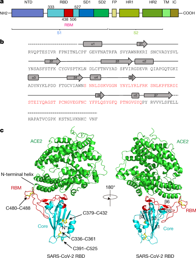
SARS-CoV-2. Specifically, we expressed the SARS-CoV-2 RBD (residues
Arg319–Phe541) (Fig. 1a, b)
and the N-terminal peptidase domain of ACE2 (residues Ser19–Asp615) in
Hi5 insect cells and purified them by Ni-NTA affinity purification and
gel filtration (Extended Data Fig. 1).
The structure of the complex was determined by molecular replacement
using the SARS-CoV RBD and ACE2 structures as search models4, and refined to a resolution of 2.45 Å with final Rwork and Rfree factors of 19.6% and 23.7%, respectively (Extended Data Fig. 2 and Extended Data Table 1).
The final model contains residues Thr333–Gly526 of the SARS-CoV-2 RBD,
residues Ser19–Asp615 of the ACE2 N-terminal peptidase domain, one
ion, four N-acetyl-β-glucosaminide (NAG) glycans linked to ACE2 Asn90, Asn322 and Asn546 and to RBD Asn343, as well as 80 water molecules.
The
SARS-CoV-2 RBD has a twisted five-stranded antiparallel β sheet (β1,
β2, β3, β4 and β7) with short connecting helices and loops that form the
core (Fig. 1b, c).
Between th
e β4 and β7 strands in the core, there is an extended
insertion containing the short β5 and β6 strands, α4 and α5 helices and
loops (Fig. 1b, c).
This extended insertion is the RBM, which contains most of the
contacting residues of SARS-CoV-2 that bind to ACE2. A total of nine
cysteine residues are found in the RBD, eight of which form four pairs
of disulfide bonds that are resolved in the final model. Among these
four pairs, three are in the core (Cys336–Cys361, Cys379–Cys432 and
Cys391–Cys525), which help to stabilize the β sheet structure (Fig. 1c); the remaining pair (Cys480–Cys488) connects the loops in the distal end of the RBM (Fig. 1c).
The N-terminal peptidase domain of ACE2 has two lobes, forming the
peptide substrate binding site between them. The extended RBM in the
SARS-CoV-2 RBD contacts the bottom side of the small lobe of ACE2, with a
concave outer surface in the RBM that accommodates the N-terminal helix
of the ACE2 (Fig. 1c). The overall structure of the SARS-CoV-2 RBD is similar to that of the SARS-CoV RBD (Extended Data Fig. 3a), with a root mean square deviation (r.m.s.d.) of 1.2 Å for 174 aligned Cα
atoms. Even in the RBM, which has more sequence variation, the overall
structure is also highly similar (r.m.s.d. of 1.3 Å) to the SARS-CoV
RBD, with only one obvious conformational change in the distal end
(Extended Data Fig. 3a).
The overall binding mode of the SARS-CoV-2 RBD to ACE2 is also nearly
identical to that observed in the previously determined structure of the
SARS-CoV RBD–ACE2 complex4 (Extended Data Fig. 3b).
The cradling of the N-terminal helix of ACE2 by the outer surface of the RBM results in a large buried surface of 1,687 Å2 (864 Å2 on the RBD and 823 Å2 on the ACE2) at the SARS-CoV-2 RBD–ACE2 interface. A highly similar buried surface of 1,699 Å2 contributed by SARS-CoV RBD (869 Å2) and ACE2 (830 Å2)
is also observed at the SARS-CoV RBD–ACE2 interface. With a distance
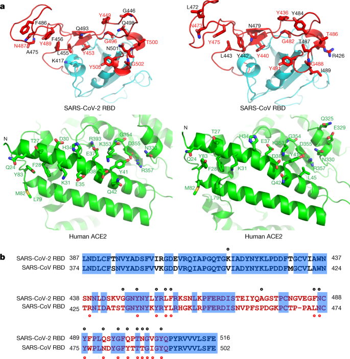
cut-off of 4 Å, a total of 17 residues of the RBD are in contact with
20 residues of ACE2 (Fig. 2a and Extended Data Table 2).
Analysis of the interface between the SARS-CoV RBD and ACE2 revealed a
total of 16 residues of the SARS-CoV RBD in contact with 20 residues of
ACE2 (Fig. 2a and Extended Data Table 2).
Among the 20 ACE2 residues that interact with the two different RBDs,
17 residues are shared between both interactions and most of the
contacting residues are located at the N-terminal helix (Fig. 2a and Extended Data Table 2).
To
compare the ACE2-interacting residues on the SARS-CoV-2 and SARS-CoV
RBDs, we used structure-guided sequence alignment and mapped them to
their respective sequences (Fig. 2b).
Among 14 shared amino acid positions used by both RBMs for the
interaction with ACE2, 8 have the identical residues between the two
RBDs, including Tyr449/Tyr436, Tyr453/Tyr440, Asn487/Asn473,
Tyr489/Tyr475, Gly496/Gly482, Thr500/Thr486, Gly502/Gly488 and
Tyr505/Tyr491 of SARS-CoV-2/SARS-CoV, respectively (Fig. 2b).
Five positions have residues that have similar biochemical properties
despite of having different side chains, including Leu455/Tyr442,
Phe456/Leu443, Phe486/Leu472, Gln493/Asn479 and Asn501/Thr487 of
SARS-CoV-2/SARS-CoV, respectively (Fig. 2b). The remaining position is at the Gln498/Tyr484 location (Fig. 2b),
at which Gln498 of SARS-CoV-2 and Tyr484 of SARS-CoV both interact with
Asp38, Tyr41, Gln42, Leu45 and Lys353 of ACE2. Among the six RBD
positions with changed residues, SARS-CoV residues Tyr442, Leu472,
Asn479 and Thr487 have previously been shown to be essential for binding
ACE218.
At the Leu455/Tyr442 position, Leu455 of SARS-CoV-2 and Tyr442 of
SARS-CoV have similar interactions with Asp30, Lys31 and His34 of ACE2
(Fig. 3a).
At the Phe486/Leu472 position, Phe486 of SARS-CoV-2 interacts with
Gln24, Leu79, Met82 and Tyr83 of ACE2, whereas Leu472 of SARS-CoV has
less interactions with Leu79 and Met82 of ACE2 (Fig. 3a).
At the Gln493/Asn479 position, Gln493 of SARS-CoV-2 interacts with
Lys31, His34 and Glu35 of ACE2 and forms a hydrogen bond with Glu35;
Asn479 of SARS-CoV interacts with only His34 of ACE2 (Fig. 3a). At the Asn501/Thr487 position, both residues have similar interactions with Tyr41, Lys353, Gly354 and Asp355 of ACE2 (Fig. 3a). Asn501 of SARS-CoV-2 and Thr487 of SARS-CoV both form a hydrogen bond with Tyr41 of ACE2 (Fig. 3a).
Outside the RBM, there is a unique ACE2-interacting residue (Lys417) in
SARS-CoV-2, which forms salt-bridge interactions with Asp30 of ACE2
(Fig. 3b). This position is replaced by a valine in the SARS-CoV RBD that fails to participate in ACE2 binding (Figs. 2b, 3b).
Furthermore, a comparison of the surface electrostatic potential also
identified a positive charged patch on the SARS-CoV-2 RBD contributed by
Lys417 that is absent on the SARS-CoV RBD (Fig. 3b).
These subtly different ACE2 interactions may contribute to the
difference in binding affinity of the SARS-CoV-2 and SARS-CoV to the
ACE2 receptor (4.7 nM compared with 31 nM, respectively) (Extended Data
Fig. 4).